


Abstract
Objective: Y-graft configuration with left and right ITA (RITA) allows complete arterial revascularisation. We previously compared two types of ITA revascularisation in a prospective randomised trial with a systematic 6-month angiographic follow-up study. The present study is a secondary analysis of these populations to evaluate the angiographic parameters influencing the function of the RITA used in a Y-graft configuration Methods: The functionality of the RITA was based on the TIMI grade flow: in TIMI grade 0 (occluded graft) and in TIMI grade 1 or 2 (balanced flow), the RITA was considered not functional. RITA was considered functional when a complete opacification (TIMI 3) of all anastomoses of the targeted coronary vessels was observed. Results: A total of 25.3% of RITA were not functional. In univariate analysis, the number of anastomoses, the type and size of grafted coronary segments and the severity of the native coronary stenosis influenced ITA function. In multivariate analysis, the function of the RITA was positively influenced by the number of anastomoses (OR = 0.5, 95% CI: 0.4–0.7), and a severely narrowed first circumflex (OR = 39.1, CI: 8.1–189.2) and negatively by the presence of a grafted intermediate coronary artery (OR = 0.01, CI: 0.003–0.06), and of a grafted RCA (OR = 0.08, CI: 0.02–0.35). The size of targeted vessel, history of infarction and regional myocardial function did not influence ITA function. Conclusions: In this systematic angiographic study, the function of the RITA used as a Y-graft was significantly improved when used on several branches of the circumflex artery or on a severely narrowed first circumflex. Grafting of the intermediate branch or of a RCA has a negative prognostic influence on graft function.
1 Introduction
Bilateral internal thoracic arteries (BITA) have clearly demonstrated their superiority over all other types of grafts in terms of patency, freedom from arteriosclerosis, and survival benefit [1,2] for revascularisation of the left coronary system.
Several configurations of BITA have been proposed to achieve complete left-sided myocardial revascularisation. Composite Y-graft configuration using the free right internal thoracic artery (RITA) graft anastomosed proximally to the left internal thoracic artery (LITA) has been widely used since its description by Barra et al. [3] and Tector et al. [4].
Concerns arose for this configuration on (a) the potential steal phenomenon of the LITA by the RITA, (b) capacity of this arrangement to revascularise completely the coronary system including the right coronary artery (RCA) [5] and (c) potential flow competition with the native coronary artery [6]. We previously addressed the first two issues by measuring the fractional flow reserve in BITA Y configuration [7]. To answer the last controversial point, we prospectively enrolled 152 consecutive patients in a systematic 6-month angiographic follow-up study to analyse the angiographic and operative parameters influencing the function of the RITA used in a Y-graft configuration.
2 Material and methods
2.1 Study design
All patients referred for isolated surgical coronary revascularisation from April 2003 to July 2006 were screened according to the inclusion criteria (Table 1 ). Patients were randomly assigned to undergo surgery according to two strategies: BITA pedicled (LITA to the LAD and RITA to the marginal branches into the transverse sinus) or BITA Y (LITA to the LAD and RITA to the marginal branches but anastomosed proximally to the LITA in a Y configuration as described by Barra et al. [3] and Tector et al. [4]. All patients were scheduled for a systematic angiography at 6 and 36 months following surgery. All patients gave written informed consent at the time of bypass surgery and before the angiographic investigation. We focused this report on the angiographic findings of the free RITA at the systematic 6-month follow-up to evaluate the preoperative and preoperative variables witch could influence the RITA function. The study protocol was approved by the ethics committee of our institution.
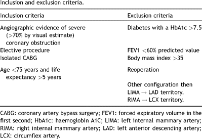
Inclusion and exclusion criteria.
2.2 Patients
From 2003 to 2006, 1297 consecutive patients underwent isolated bypass surgery in our institution. Of those, 481 (37%) patients met the inclusion criteria for study enrolment. Three hundred and four patients out of 481 (63%) were actually randomised. The remaining 177 patients were not randomised because of (a) refusal of systematic angiographic control and (b) logistic incapacity to randomise patients. BITA grafting was performed with a Y configuration in 152 (Fig. 1 ). The BITA were harvest and grafted as previously described [4–6]. The proximal composite anastomosis of the free RITA on the LITA was performed in a Y fashion after all distal anastomosis when the patient was operated with cardio pulmonary bypass. In case of off-pump surgery this anastomosis was fashioned before doing the distal anastomosis in order to protect the myocardium as the revascularisation proceeded. Operative characteristics are detailed in Table 2 . Twenty-three patients had a RITA to the PLA. Nine patients out of these had a concomitant graft to the PDA (five a saphenous vein graft and four a GEA).
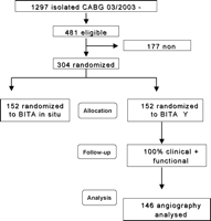
Consort flow chart of the trial.
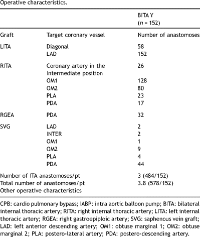
Operative characteristics.
Systematic angiographic follow-up was performed 6 months after surgery. Nitroglycerin (2 mg) was injected into each graft before filming to avoid spasm due to the catheterisation. At least two orthogonal views of each of the ITA graft were obtained, with continued exposure as required to visualise distal run-off and the size of the coronary target bed.
2.3 Data analysis
All postoperative angiograms were independently reviewed by two investigators; discrepancies in patency definition were reviewed by a third investigator and resolved by consensus. RITA function was categorised in two grades:
Not functional:
Occluded graft defined as an absence of visible opacification by the RITA of the target coronary vessel (TIMI flow grade 0 in the graft).
String sign defined as a diffusely narrowed bypass graft with a poor visualisation of the distal coronary bed.
Balanced graft defined as an anatomically patent graft with dominant flow from the native coronary or with a balanced flow supply from the native coronary and from the RITA (TIMI flow grade 1 or 2 in the graft).
Functional:
Complete opacification of all targeted coronary branches by the RITA (TIMI flow grade 3 in the graft).
Variables included in the analysis were (1) the number of distal anastomoses on the RITA, (2) the targeted coronary vessel (intermediate, circumflex 1, circumflex 2, postero-lateral artery (PLA), posterior descending artery (PDA) and the sum (PLA+ PDA), (3) the degree of stenosis of each coronary vessel (visual inspection), (4) the run-off of each targeted coronary vessel, graded from 1 to 3 according to the extent of the coronary territory beyond the stenosis on the diagnostic angiogram (Table 3), (5) the regional wall motion defined as normal, hypokinetic or akinetic on the preoperative angiogram or echocardiogram, (6) the size of the coronary artery grafted estimated by peroperative visual inspection divided into two groups 0: ≤1.5 mm and 1: >1.5 mm and (7) the history of a myocardial infarction on the regional grafted area. For the purpose of our analysis we considered as an intermediate coronary artery all coronary in the intermediate position near the left auricle. The percentage of stenosis was divided following the same groups than those described by Pevni et al. [7]: stenosis of 50% versus 51–70% versus >71%. Table 3
Run-off definition.
2.4 Statistical analysis
Data are expressed as mean ± 1 SD. In bivariate analyses, Mantel–Haenszel chi-square was used to test the associations between graft function and all binary variables.
Variables statistically significant in bivariate analyses were considered for multivariate logistic regression modelling. In a first approach, all variables that were statistically significant in bivariate analyses and that were applicable for all patients (i.e. number of anastomoses, targeted coronary artery: intermediate, first circumflex, second circumflex, posterior descending, postero-lateral and the presence of a distal RCA branch grafted (PLA + PDA)) were used. In a second approach, measures of stenosis, run-off and artery diameters were added to the first analysis when they were statistically significant in bivariate analyses and when they were applicable for more than 80% of the patients. This was the case only for CX1, which was grafted in 84% of patients. The variables analysed in the second step multivariate logistic regression modelling were the ones used for the first step added to the measures of stenosis, run-off and diameters of the first circumflex artery.
All p values are two-tailed. The Statistical Analysis Software (SAS, 9.1 releases, SAS Institute Inc., SAS Campus Drive, Cary, NC USA) was used in the statistical analysis.
3 Results
Ninety-six percent of the patients (146 patients) were controlled six months postoperatively. The remaining six patients refused the control angiography and were excluded from this study. No patient suffered from recurrence of angina during the first 6 months or demonstrated residual ischaemia during a systematic stress test done by the referring cardiologist.
From a total of 267 distal RITA anastomoses, 260 (97%) were found patent and 74.7% were fully functional according to our functional classification. Results of the bivariate analyses are shown in Table 4 . The run-off of each targeted coronary was analysed, but none of them were significantly associated with the function of the graft in bivariate analysis. The functionality of the graft was not influenced by the type of revascularisation (off-pump vs on-pump, p = 0.7).
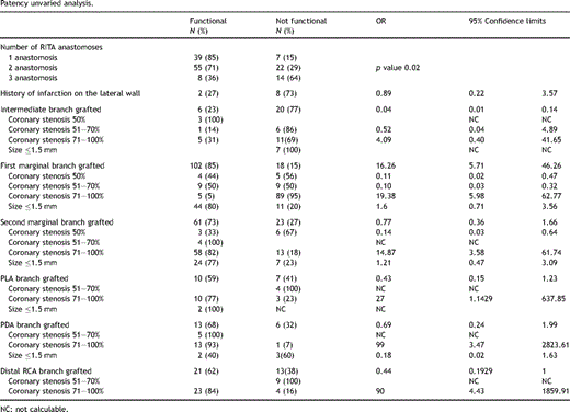
Patency unvaried analysis.
In the first step of the multivariate analysis, the function of the RITA was positively influenced by the number of anastomoses (p = 0.024) and negatively by the presence of a grafted intermediate coronary artery (p = 0.0001) and of a grafted RCA (0.0008). In the second step of the analysis, the function of the RITA was positively influenced by the presence of a grafted first marginal with a stenosis of more then 70% (0.0001) and negatively by the presence of a grafted RCA (0.0092) (Table 5 ). The size of targeted vessel, history of infarction and regional myocardial function did not influence ITA function.
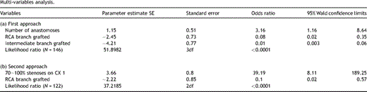
Multi-variables analysis.
The imbalance in the distribution of the intermediate coronary and right coronary grafted according to the number of anastomoses by graft might explain the inversion of the effect of the number of anastomoses on the function of the graft in the multivariable analysis.
Indeed intermediate and right coronary grafting is mainly observed where the number of anastomoses is higher. Since grafting the intermediate or the right coronary worsens the prognosis of the RITA function, the resulting effect of the number of anastomoses is reversed in the multivariable analysis: the more anastomoses the better the prognosis.
4 Discussion
This study shows that in patients treated with both mammaries in a composite Y-graft configuration, the proportion of grafts found occluded on systematic 6-month angiographic control is low. The functionality of the graft is significantly affected by the number of anastomosis, a first circumflex with a stenosis above 70%, the presence of a grafted intermediate coronary artery or a grafted RCA.
Several controversies have arisen over the BITA Y configuration. First, it is still controversial whether a single ITA can always provide sufficient blood flow, especially in the composite Y-graft to three territories. Sakaguchi et al. [8] used positron emission tomography to compare regional myocardial blood flow and flow reserve two weeks after coronary artery bypass grafting using arterial composite Y-graft or independent arterial grafts. Flow reserve was significantly lower in the Y-graft group than in the independent group, suggesting that Y-grafts are not as effective as independent graft for improving flow reserve early after coronary artery bypass grafting. Lemma et al. [9] reported the persistence of some flow reserve in the territory of composite Y-grafts during an increase in myocardial blood flow demand induced by atrial pacing early after surgery. The authors concluded that a Y-graft is able to fully respond to the flow demand of the whole left coronary system. Royse et al. [10] reported that a composite Y-graft configuration led to 75% increase of the free flow through a single ITA pedicle and that the composite Y-graft had a considerable potential of flow reserve.
Secondly, some authors have postulated that there could be a theoretical possibility of a steal phenomenon resulting in a fall of perfusion pressure in one of the branches of the Y assembling during periods of maximal myocardial blood flow demand. We have studied this phenomenon by measuring the fractional flow reserve and the pressure drops in both branches of the composite configuration at rest and during maximal hyperaemia [11]. We demonstrated that a Y-graft arterial configuration with a free right ITA attached to a pedicled left ITA allows an adequate revascularisation of the whole left coronary system with an even distribution of perfusion pressure in both distal branches and a minimal resistance to maximal blood flow. The resulting gradual decrease in pressure along the graft is negligible in basal conditions, and remains small during a maximal hyperaemia induced by coronary arteriolar vasodilation. Not surprisingly, most of the pressure drop observed during hyperaemia occurs across the common part of the Y-graft where blood flow is maximal. In addition, the conductance of both branches of the Y-graft, as assessed by FFR, appears identical, which excludes the possibility of a steal phenomenon defined as the diversion of blood flow from a high resistance to a low resistance branch during hyperaemia.
A third possible disadvantage of the BITA Y configuration is the increased risk of competitive flow in the composite graft as compared with the in situ graft. Indeed, in such assembling, the mechanism of competitive flow is more complex than that in the individual graft in which the interaction is only between the proximal inflow and the distal anastomosis outflow. In such sequential composite bypass, the interaction is also between all the anastomosed branches within the composite graft. This leads to a phasic delay between the pressure wave in the grafts, and in the coronary arteries, especially in the more distant ones such as the right coronary artery. Nakajima et al. [12] found that the most significant predictor of competitive flow and graft occlusion was the presence of a moderately stenotic branch in the RCA territory. Pevni et al. [7] issue a word of caution regarding the relatively frequent findings of competitive flow in patients with composite graft arrangement. Of major concern is their subgroup of patients with disease of the left main artery (even those with 80% stenosis) or a moderate degree of stenosis in their LAD. They concluded that composite T- or Y-grafting with two ITAs should be reserved for patients with severe stenoses in their LAD and marginal branches of the circumflex. All these findings consolidate the controversy regarding the management of moderately stenotic marginal or RCA branches. Calafiore et al. [13] have postulated that the prevention of competitive flow and graft occlusion depends on both adequate surgical strategy and manoeuvre.
We previously studied by quantitative angiography the impact of the RCA stenosis on the 6-month patency of the right gastro-epiloic artery and the saphenous vein graft [14]. We have found that a minimum lumen diameter of the RCA below 1 mm predicts a favourable flow pattern in RGEA whereas the flow pattern in SVG is significantly less influenced by quantitative angiographic parameters. We think that the RITA used in a Y-graft configuration has the same interaction with the RCA. Using quantitative coronary angiography instead of visual estimation of stenosis severity could possibly improve the value of this parameter. The absence of measurement by quantitative angiography of the left coronary system stenosis is a limitation of this sub trial, but we will focus on this in the near future.
A fourth problem with this configuration is the RITA arrangement for the intermediate branch grafting when a multiple grafting on the lateral wall of the heart is needed. Indeed we have found that grafting a coronary branch in the intermediate region had a negative prognostic influence on the RITA function. In fact, we used to systematically perform the proximal composite anastomoses of the free RITA on the LITA in a Y fashion at the posterior side (fascia) and the distal sequential circumflex anastomosis in a diamond-shape fashion. The last anastomosis of the RITA was performed in a T or T/L fashion depending on the length between this anastomosis and the last diamond-shape anastomosis. This arrangement might carry out some kinking of the intermediate anastomoses especially if the proximal Y anastomoses are performed near the pulmonary artery region or the inside of the pericardium (Fig. 2a ). Because of this finding we have changed our current practice and prefer to (a) perform a proximal T anastomoses on the LITA (Fig. 2b), (b) use a second small Y-graft to prevent seagull kinking (Fig. 2c), (c) perform the proximal composite anastomosis of a free RITA on the LITA very high on the LITA in order to obtain a smooth curve of the RITA to the intermediate branch (Fig. 2d), (d) perform a L/L anastomosis on the intermediate and a T anastomosis on the circumflex artery (Fig. 2e). These last three solutions decrease the available length of the RITA to the distal marginal branches or RCA branches.
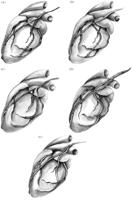
(a) Angulation of the RITA on the intermediate branch is not perpendicular; (b) proximal T anastomoses on the LITA, (c) use of a second small Y-graft; (d) proximal composite anastomosis of a free RITA on the LITA very high on the LITA and (e) latero-lateral anastomosis on the intermediate branch.
When the targeted coronary vessel in the intermediate region is the best coronary on the lateral wall, another solution would be to use the RITA in situ through the transverse sinus. In this situation we use another graft for the other smaller circumflex branches.
5 Conclusion
In this systematic angiographic study, the function of the RITA used as a Y-graft was significantly improved when used on several branches of the circumflex artery or on a severely narrowed first circumflex. Grafting of the intermediate branch or of a RCA has a negative prognostic influence on graft function.
Presented at the 22nd Annual Meeting of the European Association for Cardio-thoracic Surgery, Lisbon, Portugal, September 14–17, 2008.
This study was supported by grant no. 3.4600.04 from the ‘Fonds de la Recherche Scientifique Médicale’, Brussels, Belgium.
Appendix A
Conference discussion
Dr B. Buxton (Melbourne, Australia): I have some comments.
First of all, this group has added greatly to our understanding of the use of the right ITA as a Y-graft using functional testing rather than relying solely on anatomic studies.
The authors have a very strict classification of normal function of the right ITA graft, which has been defined as a graft which opacifies all the branches supplied by the target artery, and all should have TIMI 3 flow. Anatomically the grafts can be nonfunctional or patent as Dr Glineur has pointed out.
So the first question I would like to ask is how reproducible is this testing and the definition? Is it something anybody can use, or does it require skills to make it reproducible?In other words, the question I’m asking, what is the inter- and intra-observer error.
Dr Glineur: Well, in fact, I don’t think it's a very complex classification. We managed this – I think we were two reviewers for all the angio, myself, a surgeon, and a cardiologist. And if there was discrepancy between our point of view, then we took a third reviewer, which was a cardiologist. So it wasn’t very complex, in fact.
Dr Buxton: The next issue I have is about the statistics and the results and with the positive outcomes. They are that the greater number of anastomoses in the circumflex and high-grade stenoses in the right coronary artery associated with an excellent outcome.
Now, when you look at the univariate analysis, it seems to be reversed. The best results seem to come from the single circumflex anastomosis which is associated with a 86% patency. When you have 3 anastomoses, that drops to about 35%. I don’t quite understand this because this is in contrast to the result of the multivariate analysis.
So there is a concern. But basically the issue is that the numbers of subsets is small in the univariate analysis, and they are hard to interpret.
Now, I’m not sure who in this room is familiar with a two-stage logistic model. I suspect this is not familiar to most surgeons. Only two variables came out as being important predictors, the number of anastomoses and the target artery. But in the overall model they were not significantly predictive.
And so you took a second series of explanatory variables to see if you could define this more precisely. But by doing that, you decreased the sample size for the analysis by 20%, because by definition, they would have had an 80% or more stenosis.
So I am not sure about the relationship between the univariate and the final multivariable analysis, and it seems to be somewhat of a contradiction. Do you have any explanation for that?
Dr Glineur: Well, first of all, unfortunately, I’m not a statistician.
Secondly, I think that, of course, it's a small number of patients, and if we wanted to add more power to the statistic, we would have to control 1000 patients.
Now, I was also very surprised of the results of the multivariate analysis. I thought that it was a problem in the analysis because there was a discrepancy between the univariate and the multivariate analysis considering the number of anastomoses on the RITA. And I’ve asked that to the statistician, and he responds to me that in fact in the multiregression model, when you take out some variable, then the power of such variable can decrease, and this explained the change in the results.
But I think an explanation could be that, by increasing the number of sequential anastomosis on the RITA, you increase the run-off of the RITA. And you can think, but it's a hypothesis, that when the run-off increased, then the functionality is better. But I don’t have the real response to this.
Dr Buxton: I think we probably should just check that because the change is so large. It's from one extreme to the other.
Just another couple of comments. Next, you used the definition of minimal luminal diameter as a predictor of success in the flow of the gastroepiploic artery previously but not in the vein grafts in your current paper. Why have you not included this in this paper?
Dr Glineur: Well, of course, I was very, very interested in doing this analysis on these patients angiographic follow-up, but the problem is it's very time-consuming. And, unfortunately, I didn’t have enough time to measure all the stenoses in biplane analysis. So it's an ongoing trial that I hope I can submit next year to the European meeting, but it's very, very time-consuming.
And, moreover, it was much easier for the gastro versus the vein, to analyse this, because there was only one stenosis here. Sometimes you can – for example, if you graft three marginals, then you have to have three stenoses. And what do we do? Do we just add the percentage of MLD and divide it by three?
So it's more complex when it's sequential anastomoses than when it is just one graft, one anastomosis.
Dr Buxton: Just two follow-up quick questions. One is, what happened to the left anterior descending graft, the left internal thoracic artery to the LAD? Was that compromised by the use of the Y?
Dr Glineur: Well, we have looked at it, and there were no correlation between the RITA function and the LIMA function. It's not significant. So the LIMA is not influenced by the functioning of the RITA.
We’ve published some results three years ago in Circulation. In fact, we measured the fractional flow reserved in both arms of the Y-graft configuration. And we found that with maximal hyperaemia mimicing a stress test, we did not find any steal syndrome between both arms. So we did not find any correlation between both arms.
Dr Buxton: And perhaps one final question, when do you expect the results of the randomised trial, comparing the in situ versus the Y-graft of which this study is one arm?
Dr Glineur: They are going to be published at the end of the month in the surgical supplement of Circulation I think the 3rd of September.
Dr A. El-Essawi (Braunschweig, Germany): I have a question regarding your negative predictors. With the use of the intermediate branch, you could be too close to your Y anastomosis so you could get some kinking or something. Could you just comment if this was the reason?
And what is the reason for the worse prognosis with your branches of the RCA?
Dr Glineur: So, in fact, we wondered why did the intermediate come out, and I’ve asked one of my colleagues to do some drawings. In fact, we think that if you do your Y anastomosis too close to the intermediate artery, you can have some kinking at that site.
So we’ve proposed several other types of graft configurations. So doing a T anastomosis on the LIMA, doing a P anastomosis, put your Y to the RIMA-to-LITA anastomosis much higher or do a lateral-to-lateral, a criss-cross anastomosis on the intermediate.
But for these two solutions, the problem is that by doing this, you decrease the length that you can use of the RITA. So you can’t do, for example, the PLA or the PDA. So I think that the best configuration is to do a T anastomosis and not a Y lateral-terminal anastomosis.
This is for the intermediate for the right. Well, in fact, unfortunately, I don’t have enough controls I have something like 35 RITA to the right, distal right. The reason of poor results to the distal right is the flow competition.
Dr Buxton: I think we’re going to have to call the discussion to a close. I think we’re running over time. Perhaps if you could just finish up there.
Dr Glineur: Okay.
Dr Buxton: I think there are obviously a lot of questions unanswered by this, but it's a very provocative study.
