


PCI in a 54-year-old male with unstable angina resulted in guiding catheter-induced RCA ostium dissection, RCA obstruction and contrast retention within the aortic wall (Fig. 1 ). RCA ostium was stented and the ascending aorta replaced with Dacron graft. Postoperative CT showed aortic dissection extending to the abdominal aorta (Fig. 2 ).
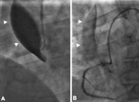
(A) Coronary angiography showing total obstruction of the right coronary artery (RCA) lumen with contrast media retention within the false lumen of the ascending aorta (arrowheads) indicative of the retrograde propagation of the RCA ostium dissection. (B) Stenting of the RCA ostium restored flow in the RCA and patient hemodynamic stability; staining of the ascending aorta wall by contrast is shown (arrowheads).
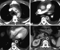
Contrast-enhanced computed tomography images demonstrate the border (arrowheads) between the true and false lumen of the dissected aortic arch (A), descending thoracic aorta (B and C) and abdominal aorta (D) at the level of the right kidney (RK) and the ostium of the right renal artery.
