


A 78-year-old man with coronary surgery 10 years ago presented with an acute onset of hoarseness and dysphagia. CT chest confirmed dissecting brachiocephalic trunk aneurysm (Fig. 1 ). The aneurysm was isolated with bypass and stenting (Fig. 2 ). Aneurysms of the brachiocephalic trunk are uncommon yet associated with incidence of high mortality.
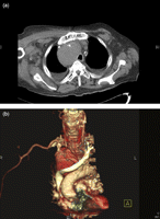
(a) CT chest at the time revealed a brachiocephalic trunk aneurysm extending from the origin of the arch of the aorta all the way up to the thoracic inlet area measuring in excess of 8 cm × 7 cm × 6 cm with evidence of surrounding thrombus and multiple lymph nodes in the pre- and peri-carinal area with the largest measuring 2.0 cm. (b) The trachea and oesophagus were compressed by the aneurysm and displaced to the left side (Fig. 2). This explained the hoarseness of voice and dysphagia for liquids and solid food in patient. The two working diagnoses were an underlying vasculitic process or a pre-existing aneurysm that became secondarily infected and rapidly expanded as per patient’s blood and urine that grew E. coli.
The aneurysm was isolated with bypass of Dacron graft and stenting (dark arrows). It was not drained at the time and 48 h later the patient had an episode of massive haemoptysis. CT scan demonstrated an aneurysmal tracheal fistula that leads to subsequent CT guided percutaneous drainage of the aneurysm. Patient recovered well.
