


A 72-year-old patient suffering from dyspnea and dysphagia. Severely elevated level of serum calcium (4.75 mmol/l). Abnormal chest X-ray. CT scan showed a 14 cm thoraco-abdominal aneurysm compressing LV and stomach (Fig. 1a ).
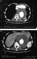
Fig. 1
CT scan demonstrates huge thoraco-abdominal aneurysm with compression of the stomach and the heart (a) and erosion of a vertebral body (b).
After aortic replacement there was normalization of hypercalcemia which was most probably caused by pressure-induced destruction of the vertebral body (Fig. 1b).
© European Association for Cardio-Thoracic Surgery 2009
