


Abstract
Objective: A prospective, randomized study to evaluate the effectiveness and safety of a polymeric sealant (Coseal®, Baxter Healthcare, Deerfield, IL) to reduce air leaks and to improve postoperative outcome in patients undergoing lung resection. Methods: Between November 2005 and February 2008, 203 (128 M, 75 F) patients showing moderate/severe intraoperative air leaks after pulmonary lobectomy/bilobectomy/sleeve lobectomy (110) or minor resection (segmentectomy/wedge) (93) have been prospectively enrolled and randomly assigned to receive one of the two following management strategies: suture/stapling (101 patients – standard care group (SCG)) or suture/stapling plus Coseal sealant (102 patients – Coseal group (CG)). To assess the effectiveness of the sealant the following data were registered and compared in the two groups: number of patients with air leak cessation intraoperatively, number of patients without air leaks at 24 h and 48 h, duration of air leaks, length of hospital stay. Results: No adverse event related to the sealant application occurred. Intraoperative air leak cessation rate was higher in the CG with a statistically significant difference (85.3% vs 59.4%; p < 0.001). Air leaks rate at 24 h and 48 h was significantly lower in the Coseal group (19.6% vs 40.6%; p = 0.001 at 24 h and 23.5% vs 41.6%; p = 0.006 at 48 h). Duration of air leaks was significantly shorter in the Coseal group (p = 0.01). The hospital stay was shorter in the Coseal group (mean: 5.7 ± 2.3 days vs 6.2 ± 2.5 days) but this difference did not reach statistical significance owing to the many known clinical interfering factors. Conclusions: The application of Coseal sealant proved safe and effective in reducing air leaks occurring after lung resection and in shortening the duration of postoperative air leak with a trend towards a shorter postoperative hospital stay.
1 Introduction
Prolonged parenchymal air leak is the most common complication after lung resection and its incidence is reported around 15–18% [1,2]. Prolonged air leak has a detrimental effect on postoperative course resulting in a longer need for chest tube with associated pain, reduced mobility and increased risk of further complications [3]. This determines prolonged hospital stay and higher costs, with an additional request for inpatient and outpatient resources [4].
Standard methods for intraoperative control of air leaks include suture and stapling, but have the disadvantage of causing further trauma to lung tissue. Various additional techniques have been employed to minimize air leaks, including the application of a number of products such as fibrin glue [5–7], synthetic sealants [8–10] and fleece bound sealants [11]. However randomized studies in this setting are rare and the results are disappointing with no clear evidence supporting the choice of an ideal technique [12].
For this reason, we have recently tested the application of a polymeric sealant (Coseal®, Baxter Healthcare, Deerfield, IL) currently employed for sealing vascular suture lines, to assess if it could play an effective role also in closing lung parenchyma.
The purpose of the present study was to evaluate the safety and efficacy of Coseal® surgical sealant for the treatment of parenchymal air leaks occurring after lung resection within the setting of a prospective randomized controlled trial.
2 Materials and methods
The trial was conducted between November 2005 and February 2008. The study population included patients older than 18 years undergoing lobectomy (including bilobectomy) or minor resections including segmentectomy and wedge resections. Patients having immune system disorders or hypersensitivity to any component of the investigational product were excluded. The trial conformed to the ethical principles of the Declaration of Helsinki and was in accordance with guidelines for Good Clinical Practice. Informed consent was obtained from each patient included in the trial before the operation.
Lung resections were performed through a lateral muscle-sparing thoracotomy. Division of incomplete fissures were performed by stapling devices (Gastro-Intestinal anastomosis 75 or 80 mm) reinforced by bovine pericardial strips. Bronchial stump suture were performed by Thoraco-Abdominal (TA) 30 mm stapler.
At the end of the surgical procedure, patients have been tested for the presence of air leaks by gradually inflating the lung with a pressure of 25 cm H2O. Air leaks, if present, have been rated subjectively as mild (countable bubbles), moderate (a stream of bubbles) or severe (coalescent bubbles) by an underwater air tightness test.
Patients with moderate to severe air leaks have been randomly allocated to one of the two following management strategies:
standard care: routine methods of closing leaks including suture or staple [standard care group (SCG)]
standard care plus Coseal: application of Coseal on air leaks sites as an addiction to the standard method (suture/staple) [Coseal group (CG)].
After the first attempt to close air leaks was performed according to the indications of each group, a second submersion test was performed to assess if the procedure was effective in closing parenchyma. In patients of the SCG the persistence or absence of the air leak was then registered without further interventions. In patients included in the Coseal group in whom air tightness was not achieved, reapplication of the sealant was performed and a third test for air tightness was conducted. The volume of Coseal applied in each patient ranged from 4 to 8 ml, depending on the extension of the surface area to be treated, the number of applications and the number of air leak sites.
Chest tubes have been regularly assessed for air leaks and fluids drainage at 24 and 48 h postoperatively by highly specialized nurses.
This study design has been chosen to best reflect routine surgical practice, where lung surgery patients who receive standard care (no sealant application) are closed up without further manipulation even if an intraoperative air leak persists. Similarly, in the Coseal arm, the possibility for a second application of the product reflects the standard mode of sealant use by surgeons.
2.1 Materials
Coseal surgical sealant (Baxter Healthcare, Deerfield, IL) is a sprayable polymeric matrix. Its basic mechanism is the rapid formation of a biocompatible hydrogel that firmly adheres to tissue and thereby provides rapid sealing of the site. The hydrogel is resorbed by the body within 30 days from application.
The primary aim of this study was to assess if the Coseal was able to increase the proportion of patients showing air leak cessation intraoperatively (no air leak at the second or third air tightness test).
The secondary aim was to assess if Coseal was able to improve postoperative outcome reducing: the number of patients with air leak at 24 and 48 h after surgery, the duration of air leak, the length of hospital stay, the need for intensive care unit (ICU) stay and the associated morbidity.
Moreover, another purpose of the study was the assessment of the safety of the investigated product by the evaluation of the incidence of adverse events.
2.2 Randomization
Entry to the trial and randomization took place in the operating room once it was established that a moderate or severe air leak persisted following the standard surgical procedure. The randomization sequence has been generated by the Medical Informatics, Biometry, and Epidemiology, University of Munich (Germany) and included in a series of sealed envelopes provided to the investigators. The operating surgeon ascertained the treatment allocation for each eligible patient by opening the next available sealed randomization envelope of this series.
2.3 Statistical analysis
Descriptive statistics have been applied to primary and secondary outcome measures for all the patients. Percentage rates have been compared between the two treatment groups by the chi square test.
Quantitative measurements have been compared between the two groups using the Mann–Whitney test. Differences have been considered significant if p was ≤0.05.
Continuous data are presented as mean ± standard deviation.
Confidence intervals (95%) were used to quantify the extent of the observed differences.
3 Results
Overall 203 patients fulfilled the inclusion criteria at the end of lung resection and were enrolled into the study. One hundred and twenty-eight (63.1%) were male and 75 (36.9%) female with age ranging between 30 and 83 years (mean 57.5 ± 12.3). Of these patients, 101 (67 males, 34 females; age range 30–80 years, mean 58.2 ± 12.2) were randomly assigned to the SCG and 102 (61 males and 41 females; age range 34–83 years, mean 56.8 ± 12.5) to the CG. Patients’ characteristics according to gender, age, height, weight and body mass index (BMI) did not show significant differences between the two groups (p > 0.05). The distribution of patients according to the presence of smoking history, chronic obstructive pulmonary disease (COPD) and abnormal preoperative and predicted postoperative FEV1 was similar between the two groups as well (Table 1 ).
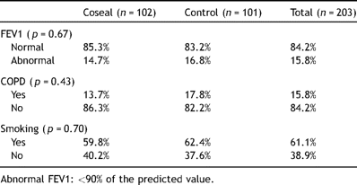
Preoperative FEV1/comorbidities.
Surgical procedures performed in the 203 patients included 104 lobectomies, 6 bilobectomies, 2 sleeve lobectomies and 91 lesser resections (12 segmentectomies, 79 wedge resections). The distribution of the surgical procedures in the two groups is reported in detail in Table 2 and it did not show any significant difference (p = 0.17).

Surgical procedures.
In the CG the dose of Coseal application after the first underwater air tightness test was 4 ml in 96 patients (94.1%) and 8 ml in 6 patients (5.9%). After the second air tightness test the persistence of air leak was observed in 23 patients of the CG and a second application of 4 ml of Coseal was performed in these cases.
Intraoperative air leak cessation was obtained in 60 patients (59.4%) of the SCG and in 87 patients (85.3%) in the CG. This difference was statistically significant (p < 0.001, 95% CI [14.1%, 37.7%]). At 24 h postoperative assessment, air leak was present in 41 patients (40.6%) of the SCG and in 20 patients (19.6%) of the CG, with this difference being statistically significant (p = 0.001, 95% CI [−33.3%, −8.7%]). At 48 h postoperative evaluation 42 patients (41.6%) in the SCG and 24 patients (23.5%) in the CG presented an air leak. This difference was also statistically significant (p = 0.006, 95% CI [−30.7%, −5.4%]).
The duration of air leaks was significantly shorter in the CG (mean 3.5 ± 1.7 days; range 1–10) with respect to the SCG (mean 4.2 ± 2.4 days; range 1–14) (p = 0.01, 95% CI [0.13, 1.27]).
There was a trend towards shorter hospital stay in the CG (mean 5.7 ± 2.3, median 5; range 2–13 days) with respect to the SCG (mean 6.2 ± 2.5, median 6; range 2–16 days), but this difference was not statistically significant (p = 0.18).
The occurrence of associated postoperative complications was registered in three patients (3%) in the SCG and in two patients (2%) in the CG with no statistically significant difference between the two groups (p = 0.64). Associated complications are reported in details in Table 3 . There was no perioperative mortality.
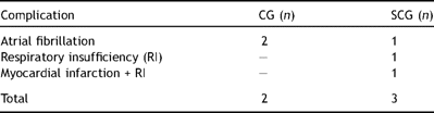
Complications.
Intensive care unit (ICU) stay was necessary in two patients (2%) in the SCG and in two patients (2%) in the CG (p = 0.99).
The volume of chest tube drainage (pleural fluids) up to the second postoperative day did not show a statistically significant difference (p = 0.81) between the two groups (SCG: mean volume 386.9 ± 184.0 cc; CG: mean volume 393.7 ± 205.6 cc).
4 Discussion
Although significant efforts have been made to reduce the incidence of parenchymal air leaks following lung resection, including the search for improved surgical technique and the experimentation of a number of sealants, an ideal treatment for this surgical complication has still not been identified.
Intra-operative air leaks after standard pulmonary resections are reported ranging between 48% and 70% [9,11] in main series, and prolonged air leaks persisting over 7 days have been observed in 15–18% of cases [1,2].
The risk increases when interlobar fissures are incomplete and in patients with emphysematous lung. Moreover, the presence of reduced predicted postoperative forced expiratory volume at 1 s (ppo-FEV1), pleural adhesions and upper resections have been found to be predictors of prolonged air leaks in large retrospective studies [1].
The routine use of surgical staplers for division of parenchyma has improved the primary closure of resection lines. Buttressing of staple line with synthetic or biological materials including bovine pericardial strips [13] has been recommended based on the positive results achieved in emphysema surgery. However, this technique can only be used in a prophylactic setting, since a number of further potential sources of air leak have to be considered, especially in the hilar region and on decorticated lung surface areas.
For this reason increasing requirements for new sealant products to be used as an adjuvant to surgical technique has stimulated active industrial research and clinical experimentation in this setting.
Fibrin glue has been the most studied option over the last two decades, unfortunately with unsatisfactory results. The randomized trials conducted by Wong and Goldstraw [5] and by Fleisher and colleagues [6] have demonstrated no difference in the incidence of postoperative air leaks, in the duration of chest-drain and in hospitalization.
In contrast, a recent randomized study analysing the effects of an autologous fibrin sealant [14] has shown a significantly reduced proportion of patients with air leaks and lower mean air leak volume in the arm treated with the product. However, the small size of the series analysed in this study may represent a limit to the power of its results.
Recently, three randomized trials [8–10] have tested the efficacy of synthetic bio-absorbable (polyethylene glycol-based) sealants, observing a significantly lower air leak rate with a trend towards shorter chest-drain duration and hospitalization in the treated group, although significant difference in the latter two parameters was not found. However, an increased incidence of postoperative empyema has been registered in one of this studies [8], suggesting the possibility of insufficient adhesion of the sealant to the lung in some cases, determining an action as foreign body into the pleural cavity.
Current clinical experimentation in this field has indicated that sealant materials have to display a number of characteristics. They need to be sufficiently adherent to withstand pressure of the inflated lung, but also elastic to accommodate the volume changes of the parenchyma during respiration. Moreover these materials should also fix rapidly to the lung tissue and be unaltered by the intrapleural fluids. They also need to be nonirritating, systematically non-toxic, and without antigenicity.
We have found many of these characteristics in the Coseal sealant whose basic mechanism is the rapid formation of a biocompatible hydrogel that firmly adheres to the tissue. This evidence and the positive results observed in our previous study with this sealant [15] have induced us to test the product in a larger, adequately powered, prospective randomized trial.
The application of this synthetic sealant in the present experience has allowed the significant reduction of a proportion of patients showing intraoperative and postoperative air leaks assessed 24 and 48 h after surgery.
Furthermore, among the patients who received sealant therapy but showed postoperative air leaks, the duration of air leaks was significantly shorter if compared with the standard care group.
There was a small number of patients (5) in the CG showing reappearance of air leak after initial postoperative evidence (at 24 and/or 48 h) of effective air tightness. Revision of these patients’ records has allowed us to establish that in almost all of them multiple air leak sites were visible after resection requiring the division of the standard quantity of sealant in smaller doses for each parenchymal area to be treated. This evidence suggests the need for increasing the dose of the sealant in patients with multiple sites of air leaks.
Despite the clinical advantage obtained by the earlier air leak cessation, a statistically significant reduction in the duration of the hospital stay was not found in patients receiving sealant application although they showed a favorable trend towards shorter hospitalization. This result should not be a surprise because air leaks are not the only major reason for postoperative hospitalization. Many other medical and surgical factors may influence patient recovery and have an impact on hospital stay.
We have not observed any specific complication related to the use of the sealant and postoperative morbidity was similar in the two groups. Moreover the sealant application did not lead to increased volume of chest tube drainage or pleural effusion.
In conclusion, the application of Coseal sealant in this large randomized study proved safe and effective in reducing air leaks occurring after lung resections and in shortening the duration of air leak with a trend towards a shorter postoperative hospital stay.
Presented at the 22nd Annual Meeting of the European Association for Cardio-thoracic Surgery, Lisbon, Portugal, September 14–17, 2008.
Dr Claudio Andreetti is the recipient of a grant financed by Baxter Healthcare.
