


Abstract
Objective: Optimising the preservation modality and thus maintaining the post-transplanted organ function remains a point of interest in research in order to prevent deleterious ischaemia/reperfusion injury. Microcirculation allows the assessment of initial graft function before obvious functional parameters. It was the aim of our study to compare the effects of epoprostenol and sildenafil on the pulmonary microcirculation and haemodynamics, when used in the preservation solution in lung transplantation. Methods: Twenty-one pigs underwent single LuTx after 24 h graft-ischaemia, preserved with buffered low potassium-dextran solution (I, control); with addition of 0.66 μg/kg/bw epoprostenol (II) or with 0.15 mg/kg/bw sildenafil (III). The pulmonary microcirculation, alveolar capillary diameter (ACD), red blood cell (RBC) velocity and functional capillary density (FCD), were assessed by intravital microscopy (OPS-imaging) hourly until 6 h after reperfusion. Haemodynamics and blood gas exchange were monitored at all timepoints. Results: ACD was increased in group III directly after reperfusion (132 ± 4.4% vs 121 ± 3.1%, in % of baseline, III vs I; mean ± SEM; p < 0.05) and decreased during the experiment. RBC velocity did not reach statistical significance (256 ± 93 vs 263 ± 85 and 283 ± 66 μm/s, III vs II and I; mean ± SD). FCD in group III was higher than in I and II beginning 3 h after reperfusion (10.1 ± 1.4 vs 6.1 ± 1.9 μm/μm2, III vs I; mean ± SEM; p < 0.05). Conclusions: Our study demonstrated a significantly improved microcirculation after application of PDF V during organ procurement, probably because of better distribution of the preservation solution. Further studies are necessary, to prove the long-term effects of this observation.
1 Introduction
Lung transplantation has evolved to a recognised therapy of terminal end-stage lung disease. However, postoperative dysfunction with imminent association to graft failure and/or impaired long-term function still impairs the clinical results of this procedure [25].
These complications are usually attributed to alterations of the lung in its cellular and functional entity due to the multifactorial ischaemia-reperfusion (IR) injury [9,19].
Besides other reasons a resulting severed microcirculation might be the reason of early postoperative disorder.
In solid organ transplantation different animal models have investigated the role of the microcirculation, the deleterious consequences of its alteration and the possible interventions in order to prevent/improve the microcirculatory disorder with promising results [2,16,20–23].
In vivo visualisation of the organ microcirculation has been limited due to the obvious large sized technical setting and the need of fluorescent dyes. The recently established orthogonal polarisation spectral (OPS) imaging allows in vivo, direct visualisation and quantification of the organ microcirculation without the use of fluorescent dyes with reliable results in experimental comparative studies [12,14,21–23]. With the help of this promising technique, microcirculation changes and the effect of novel modalities in organ preservation can be observed.
Various experimental setups demonstrated microcirculatory disturbances in solid organ transplantation correlating with tissue damage as well as functional impairment. Furthermore the degree of microcirculation disturbance could reflect on the tissue injury and therefore allow for early medical intervention with subsequent benefit on graft survival and overall morbidity [22,11]. In organ retrieval microcirculation could even offer additional information helping the surgeon decide if the organ qualifies for transplantation [26]. In lung transplantation the microcirculation disorder has been associated with the development of rejection and/or the bronchiolitis obliterans syndrome (BOS), an observation of particular relevance since tissue perfusion is compromised postoperatively because of the often not reanastomosed bronchial artery [1,3].
In previous studies we have investigated the use of the phophodiesterase-5 (PDE-5) inhibitor sildenafil during preservation in an experimental lung transplantation model with promising results [19]. In this work we observed a beneficial effect when sildenafil was applied into the pulmonary trunk whereas sildenafil in the preservation solution led to an increased water content of the transplanted lung with functional restrictions. Therefore we decided to pursue the first model of application with direct injection of sildenafil into the pulmonary artery; additionally, after 20 min the effect of sildenafil was limited on the pulmonary circulation in contrast to longer lasting systemic effects observed after using epoprostenol [8,24,27]. Due to its high selectivity, sildenafil already has demonstrated a superior positive effect regarding post-transplanted lung function when compared to the clinically used epoprostenol, using low potassium-dextran (LPD) solution as preservation solution in the work of Korom et al. [10]. In this study from 2006 epoprostenol was administered into the pulmonary artery and led to severe lung oedema 2–3 h after transplantation. Considering these results, we decided to use epoprostenol only as additive in the preservation solution as in our present clinical standard.
Therefore it was the aim of our study to compare the effects of epoprostenol and sildenafil on the pulmonary microcirculation and macro-haemodynamics, when used with the preservation solution LPD in lung transplantation. Additionally, we wanted to assess the microcirculation and its possible use as post-transplant function parameter.
2 Materials and methods
The following protocol was reviewed and approved by the committee on research animal care, Essen Medical School. All animals received humane care in compliance with the Principles of Laboratory and Animal Care and the Guide for the Care and Use of Laboratory animals prepared by the Institute of Laboratory Animal Resources published by the National Institutes of Health (NIH publication no. 85-23, revised 1985).
2.1 Experimental groups and animals
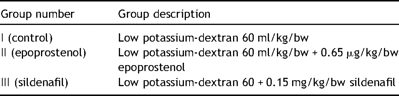
Forty-two male pigs, German landrace (30–35 kg), were included in this study, 21 serving as donor animals and 21 as recipients. Donor animals received antegrade flush perfusion (0.5 l/min) with TRIS-buffered low potassium-dextran solution (LPD; Vitrolife, Göteburg, Sweden) (group I, control, n = 7), with addition of 0.6 μg/kg bw epoprostenol (Flolan®) (group II, n = 7) and after intravenous application of 0.15 mg/kg bw sildenafil directly in the pulmonal artery trunk (group III, n = 7) (Table 1 ). The retrieved grafts were stored for 24 h at 4–6 °C and transplanted as single left lung.
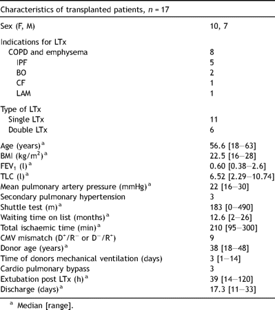
Test group description.
2.2 Surgical procedures
In this study, we used our model of left-sided lung transplantation that has been described in detail previously [14]. Shortly, anaesthesia was performed with disoprivan (10 mg/h), midazolam (1 μg/kg/h) and fentanyl (5 μg/kg/h) continuously administered intravenously. Mechanical ventilation (Fabius GS, Dräger, Germany) was performed in a pressure-controlled mode with standard settings: ventilation rate of 15 breaths/min, maximum inspiratory pressure of 25 mmHg, positive end-expiratory pressure 5 mmHg at an inspiratory oxygen-tension (FiO2) of 0.5 at explantation and 1.0 at implantation. ECG was connected and a catheter was placed in the femoral artery for haemodynamical monitoring and later blood gas analysis in all animals.
2.2.1 Donor operation
After median sternotomy a flushing tube was inserted into the main pulmonary artery. Systemic anticoagulation was performed with 3 mg/kg/bw heparin. Under inflow occlusion, the aorta was clamped and flush perfusion with 60 ml/kg bw of 4–6 °C cold-buffered LPD solution was induced. In group II the buffered LPD solution was supplemented with 0.6 μg/kg bw epoprostenol. In group III 0.15 mg/kg bw sildenafil was injected directly in the pulmonal artery trunk prior to flush perfusion. Ventilation of lungs was maintained throughout the entire procedure. The lungs were excised en bloc and stored mildly inflated in a refrigerator for 24 h in 4–6 °C cold LPD solution.
2.2.2 Recipient operation
In addition to the standard monitoring, a Swan–Ganz catheter (Baxter, Healthcare Corp., Irvine, CA) was placed through a jugular vein introducer sheath for complete haemodynamic monitoring.
After left-sided lateral thoracotomy the tracheal bifurcation, the left pulmonary artery and the left pulmonary veins were exposed. Heparin (3 mg/kg) was administered intravenously. After clamping of the left main bronchus and the left pulmonary artery, the main stems of left pulmonary veins were ligated. Pneumectomy was performed. The left atrium was clamped. The donor lung was implanted in a typical manner with an end-to-end anastomosis of the left bronchus, left pulmonary artery followed by the atrial cuff combining the left pulmonary veins. After deairing the pulmonary artery was declamped and the graft was ventilated. After a reperfusion period of 10–20 min the contralateral pulmonary artery and bronchus were clamped, in order to evaluate only the function of the transplanted lung. If necessary, haemodynamic support was applied by administration of epinephrine or noradrenaline (maximum 0.4 μg/kg bw/min). Each experiment was terminated after an observation period of 6 h by an intracardial tetracainchloride (T61) injection.
2.3 Monitoring and measurements
All animals were evaluated before procedure (baseline), at 30 min, 1 h, 3 h and 6 h after transplantation.
2.3.1 Haemodynamics and lung function
Haemodynamic data were monitored continuously (Siemens SC 9000 and Swan–Ganz catheter, Baxter). Arterial blood gas analysis (Radiometer Copenhagen, Germany) and circulatory parameters (cardiac output (CO), pulmonary vascular resistance (PVR) and systemic vascular resistance (SVR)) were performed at each timepoint. The system was calibrated by injection of cold saline solution via the Swan–Ganz catheter and parameters were calculated by the monitor system.
2.3.2 Microcirculation assessment
2.3.2.1 Technique of OPS imaging
Linearly polarised light is emitted from the objective probe into the tissue. A second orthogonally oriented polariser is used to block light reflected from tissue surfaces with unchanged polarisation before visualisation. Light which is multiply deflected and scattered in deeper layers of the tissue experiences a change of its polarisation direction. This light passes the second polariser and serves as a virtual light source in the depth of the tissue. Thus, images comparable to those obtained by transillumination are created.
The technique of OPS imaging has been described in detail previously by the working groups of Groner et al. and Lindert et al. [5,12].
2.3.2.2 Cytoscan
OPS technique illuminates the tissue with light of a wavelength of 548 nm, which is, absorbed equally well by oxy- and desoxyhaemoglobin to improve visualisation of blood vessels. With a 10× probe (334× magnification on the 10.4-in. display), the resolution is about 1.0 μm and thus high enough to allow differentiation of single erythrocytes in capillaries and intravascular optical patterns created by erythrocytes and plasma gaps in larger vessels.
In our setting, OPS images were obtained from standardised areas of the lung surface. In order to avoid direct contact and the application of pressure to the tissue area under observation we used a modified distancing/immobilising device as described by Lindert et al. [12].
Three sequences of motion pictures (50 frames, with a frequency of 30 frames/s) were recorded at each timepoint and stored on the Cytoscan® hard drive. Later, data were transmitted to a PC-workstation for off-line analysis [6].
Images were digitalised by means of a frame-to-frame computer-assisted image analysis system, CapiScope® (KKtechnology, Devon, United Kingdom). Microvessel diameters, red blood cell (RBC) velocity, and functional capillary density (FCD) were measured. FCD is defined as the length of RBC – perfused capillaries (4–12 μm diameter) per observation unit area and is given as μm/μm2.
2.4 Statistical analysis
2.4.1 Haemodynamical data
All data were expressed as mean ± standard deviation (SD). Data were analysed by repeated measure analysis of variance (ANOVA) and paired t-test. For data without repeated measurements a Mann–Whitney U-test was performed; p values less then 0.05 were considered significant.
2.4.2 Microcirculation data
All data were expressed as mean ± standard deviation of the mean (SEM). After passing the normality (Kolmogorov–Smirnov) test differences were analyzed by ANOVA for repeated measures followed by post-hoc Bonferroni correction; p values less then 0.05 were considered significant.
All data were analysed and represented using SigmaStat version 3.5.0 (Systat Software GmbH, Erkrath, Germany) and Microsoft Office Excel 2003 (Microsoft Corporation, USA).
3 Results
3.1 Animal survival
All animals of the three groups (n = 7 in each group) survived during the whole observation period.
3.2 Haemodynamical data
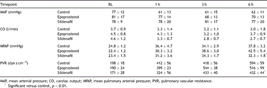
All groups showed similar general haemodynamic values and a stable heart function (Table 2 ).
Haemodynamic data, n = 7. All values are mean ± SD.
Mean PAP was elevated after transplantation in all groups, however group III showed significant lower values at timepoint 6 h when compared to control (p < 0.01) (Table 2).
PVR was similarly elevated in all groups after transplantation, but group III presented significant lower values 6 h after reperfusion when compared to control group (p < 0.01) (Table 2).
3.3 Pulmonary gas exchange data
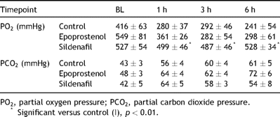
Oxygenation capacity of the transplanted lung was significant higher in the sildenafil group at all timepoints after reperfusion when compared to control group (p < 0.05) (Table 3 ).
Arterial blood gas results, n = 7. All values are mean ± SD.
Arterial partial carbon dioxide fraction was elevated after reperfusion and showed no significant difference between the different groups, although group II showed a trend of higher fractions as shown by the mean values (Table 3).
3.4 Microcirculatory data
Mean 345 ± 121 vessels were assessed at each timepoint and in each group during evaluation of the microcirculatory parameters.
3.4.1 Alveolar capillary diameter
Alveolar microvessel diameter showed a distinct increase after transplantation in each group until end of observation time. Group III presented with a significant higher mean diameter when compared to group I at timepoint 30 min (p < 0.01) but showed regression during the observation time. Group I and group II stayed on similar levels as direct postoperatively (Fig. 1 ).
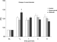
Percentual change of pulmonary microvessel diameter in comparison to the baseline given in % (all values are mean ± SEM, * significant vs control, p < 0.01).
3.4.2 RBC velocity
RBC velocity showed no significant differences after transplantation until end of experiment (Fig. 2 ). Values were overall lower in control group and group II, showing a recovery until timepoint 6 h, whereas group III presented with stable values comparable with baseline.
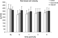
RBC velocity in pulmonary microvessel given in μm/s, n = 7. All values are mean ± SEM. No significance was achieved.
3.4.3 Functional capillary density
FCD showed distinct lower values in all groups after transplantation. Group III showed a significant recovery 3 h after transplantation with significant higher values at timepoint 3 h and 6 h when compared to control (p < 0.01). All groups showed a trend to increasing FCD during the observation time.
3.4.4 Wet/dry ratio
Lung water content showed no statistical differences in the different groups. However a slight increase in the sildenafil group (5.6 ± 0.9 vs 4.3 ± 0.9 and 4.8 ± 0.8, epoprostenol and control, respectively) was noted.
4 Discussion
In the present study we demonstrated a recovery of the functional capillary density with significant better values in the PDE-5 inhibitor treated group in correlation with improved functional parameters like oxygenation ratio and pulmonary vascular resistance.
These findings go along with studies implying the importance of the FCD in the context of organ function.
The microcirculatory changes after lung transplantation are mainly expressed as a dilatation of the pulmonary microvessels and a reduced functional capillary density with only slow recovery in the defined observation time of 6 h. These findings are comparable with observations in other transplanted organs as liver and pancreas [1,22,23], analogue to the ‘no reflow’ effect [15]. Furthermore, the degree of microcirculation disturbance after reperfusion is postulated to correlate with the early graft function and recovery [18,23]. The PDE-5 inhibitor sildenafil leads to an increased vasodilatation 30 min after transplantation, but showed a consistent recovery when considering the diameter of group I and II, which did not change within 6 h of transplantation (Fig. 1). FCD showed excellent recovery in the sildenafil group with almost 75% of the baseline values at timepoint 6 h (Fig. 3 ), demonstrating a superior protective effect when compared to epoprostenol. These results are supported by the clinical data collected in this setting: improved oxygen capacity, lower mean arterial pressure and pulmonary vascular resistance, all factors known to be indicators of good initial and long-term lung graft function [17,19].
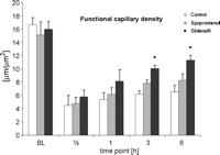
Functional capillary density (FCD) given in μm/μm2, n = 7. All values are mean ± SEM, * significant versus control, p < 0.01.
Pulmonary vasodilatators like epoprostenol, a prostacycline derivate, are used as additives in order to counteract reactive vasoconstriction in the graft of brain death donors [7,9] and thus permit a complete homogenous perfusion leading to optimal preservation.
The newer agent sildenafil is a potent selective phophodiesterase-5 inhibitor and is increasingly used in clinical settings [4,13,24,27]. The vasodilatative effects of sildenafil are due to accumulation of cGMP, inducing vasodilatation especially in lung tissue because of its high occurrence of the phosphodiesterase-5 subtype, resulting in less significant systemic effects than the clinically used epoprostenol [24,27].
The observed increase of the pulmonal microvascular diameter and of the functional capillary density could be the combined result of a better protection and a continued vasodilatation leading to opening of shunted capillaries, which allow for a higher blood circulation in the lung. This increased blood flow, which could not be reached by the non-specific vasodilatator epoprostenol, can be interpreted as a direct result of the PDE-5 inhibitor during explantation and therefore explain the superior postoperative graft function after 24 h of cold ischaemia. However, wet/dry ratio indicated a trend to higher values in the sildenafil group without reaching significance (Table 4 ), leading to the worry of potential oedema formation in the lung depending on the extent of microcirculation effects of sildenafil. Dosage and/or length of ischaemia may play a role and should be evaluated further.
Wet/dry ratio in different groups, n = 7, all values are mean ± SD; no significance between groups was achieved.
In conclusion, our experiments demonstrate an improved function of the transplanted lung graft due to a better microcirculatory perfusion after PDE-5 inhibitor modified preservation when compared to the clinically used epoprostenol. OPS-imaging was found to be a valuable tool in assessing in vivo the microcirculation of the lung, providing data consistent with the usual clinical parameters. However more investigations are needed to further understand the role and meaning of the disturbed post-transplanted lung microcirculation.
Acknowledgments
The authors would like to thank Mrs Karin Kaiser for her technical assistance during the experimental procedures as well as Professor Ghazwan Butrous for his council.
