-
PDF
- Split View
-
Views
-
Cite
Cite
Hui-li Gan, Jian-quan Zhang, Occupying the whole pulmonary artery: chronic thromboembolic pulmonary hypertension, European Journal of Cardio-Thoracic Surgery, Volume 35, Issue 4, April 2009, Page 725, https://doi.org/10.1016/j.ejcts.2009.01.002
Close - Share Icon Share
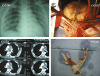
An adult woman presented with faints and shortness of breath. X-ray revealed a filling defect in the pulmonary arteries (Fig. 1A and B ). A thrombus was removed during pulmonary thromboendarterectomy procedure (Fig. 1C and D). The systolic pulmonary artery pressure decreased from preoperative 115 mmHg to 45 mmHg with a 6-month follow-up.

(A) The chest röntgenography. (B) The pulmonary artery CT angiography (transverse view) showed a whole filling defects in the main and both of the right and left pulmonary artery. (C) The thromboendarterectomy procedure. (D) An enlarged and organized thrombosis which extended from the pulmonary valve to both the right and left pulmonary artery and their branches was removed.


