-
PDF
- Split View
-
Views
-
Cite
Cite
Francesco Leo, Domenico Galetta, Alessandro Borri, Lorenzo Spaggiari, Segmentectomy for carcinoid arising from an accessory cardiac bronchus, European Journal of Cardio-Thoracic Surgery, Volume 35, Issue 3, March 2009, Page 537, https://doi.org/10.1016/j.ejcts.2008.12.015
Close - Share Icon Share
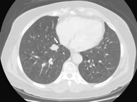
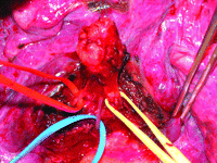
A right lower lobe nodule associated with 2 mm nodule of the middle lobe was discovered in a 74-year-old female. The major lesion was in an accessory cardiac bronchus (Fig. 1 ) which was anatomically removed for diagnostic purpose (Fig. 2 ). Both lesions were typical carcinoids. This abnormality has never been reported before.

At CT scan, the typical aerated appearance of the accessory cardiac bronchus was not present due to proximal mechanical obstruction. Its presence may be only presumed by the pleural folding sign (arrow). Bronchoscopy, performed for the presence of hemoptysis, did not reveal any anatomical abnormality. Diagnosis of accessory cardiac bronchus was made only intra-operatively.

Anatomical structures of the accessory cardiac bronchus were isolated and separately ligated (artery: red, vein: blue, bronchus: yellow). Given the metastatic nature of the middle lobe nodule and the patient’s poor respiratory function, bilobectomy was not performed. Two years after surgery, no recurrence was recorded.


