-
PDF
- Split View
-
Views
-
Cite
Cite
Mario Santini, Alfonso Fiorello, Luigi Mansi, Pier Francesco Rambaldi, Giovanni Vicidomini, Luigi Busiello, Gaetana Messina, Paola Nargi, The role of technetium-99m hexakis-2-methoxyisobutyl isonitrile in the detection of neoplastic lung lesions, European Journal of Cardio-Thoracic Surgery, Volume 35, Issue 2, February 2009, Pages 324–331, https://doi.org/10.1016/j.ejcts.2008.09.033
Close - Share Icon Share
Abstract
Objective: Our goal was to determine the role of technetium-99m hexakis-2-methoxyisobutyl isonitrile (99mTc-MIBI) in the detection of neoplastic lung lesions. Materials and methods: We prospectively studied 79 consecutive patients with indeterminate lung lesion between January 2006 and September 2007. All patients were submitted to 99mTc-MIBI single-photon emission chest tomography (SPECT) before invasive diagnostic procedure. Qualitative analysis was performed to evaluate SPECT images in order to localize abnormal activity in the radiologically demonstrated lesion. In addition, semiquantitative analysis was made by calculating tumor/contralateral normal lung ratio (T/N). Finally, the scintigraphic findings were correlated to the histopathological diagnosis obtained by invasive procedure or confirmation of instrumental exams. Results: Sixty patients had a malignant lesion: 44 squamous cell carcinoma, 7 adenocarcinomas, 4 large cell carcinoma, 1 small cell lung cancer, and 4 metastases. The mean size ± standard deviation of malignant nodules was 3.9 ± 1.61 cm (range 1.5–5.5 cm). Nineteen patients had a benign disease. The mean size ± standard deviation of benign nodules was 3.3 ± 1.71 cm (range 2–6 cm). 99mTc-MIBI SPECT delineated focal lesions with an increase in tracer accumulation in 55/60 malignant lesions; in 5/60 malignant lesions was negative. Sensitivity, specificity, positive predictive value (PPV), and negative predictive value (NPV) were 91%, 73%, 91%, and 73%, respectively. In patients with neoplastic lesion, the mean T/N ratio value ± standard deviation was 1.72 ± 0.35 whereas in patients with benign lesions was 1.14 ± 0.25. Semiquantitative analysis showed that for a T/N value >1.23, the value of sensitivity, specificity, PPV, and PNV were 91%, 84%, 94%, and 76%, respectively (ROC curve). Metastatic mediastinal lymph nodes were found in 3/57 patients. 99mTc-MIBI SPECT showed a specificity and PPV of 100% in the detection of mediastinal lymph nodes with sensitivity, and PNV of 66% and 97%, respectively. Age, sex, histological type, and size of lesion did not affect the SPECT results. Conclusion: Our experiences seem to confirm that 99mTc-MIBI SPECT is a reliable diagnostic tool in the finding of lung cancer particularly cases in which radiological evaluation is indeterminate.
1 Introduction
Pulmonary nodules and masses frequently present a diagnostic dilemma (benign or malignant lesion?) especially when they are solitary, surrounded by normal lung tissue, and not greater than 3.0 cm in its largest diameter without radiographic evidence of hilar or mediastinal adenopathy (solitary pulmonary nodules). Non-invasive testing results from patients with suspected lung cancer are frequently unreliable. Conventional imaging techniques have a limited diagnostic accuracy since interpretation relies principally on lesion size and other non-specific findings [1]. Definitive diagnoses have traditionally depended on invasive techniques. However, even invasive procedures such as bronchoscopy and transbronchial or transthoracic biopsy have sensitivities of ≪80% in certain settings and may be associated with significant complications [2]. For these reasons, newer imaging methods that do not rely solely on lesion size have been considered in patients with lung cancer. There is thus an increased interest in the functional non-invasive radioisotopic methods in attempting to reduce the number of invasive diagnostic procedures. These methods, which demonstrate the metabolic properties of a lesion, are particularly indicated in differentiating benign from malignant forms especially in patients with indeterminate pulmonary mass [3]. F-18–2-fluoro-2-deoxyglucose (FDG) positron emission tomography (PET) is one such examination that takes advantage of increased glucose utilization by tumor cells [4,5]. However, FDG PET has some limits. First, a variety of inflammatory lesions (tuberculosis, sarcoidosis, inflammatory pseudotumor, fungal infection, pneumonia, and abscess) have all been associated with FDG PET uptake [6]. Second, adenocarcinoma and bronchioloalveolar lung carcinoma may be not visualized by FDG PET [7]. Third, FDG PET remains an expensive and complicated procedure, which is performed with tracers and imaging equipment that are available at a limited number of sites. Consequently, various radio nuclides, such as 67Ga, 201Tl, and Tc-99m tetrofosmin have been utilized in lung cancer for staging [8–10], follow-up, and monitoring the response to therapy [11]. In addition, encouraging results have also been obtained with single-photon emission chest tomography (SPECT) scanning using technetium-99m hexakis-2-methoxyisobutyl isonitrile (99mTc-MIBI). 99mTc-MIBI is a lipophilic cation widely used as a tracer for myocardial perfusion imaging but its usefulness has been demonstrated in particular in tumors sited in the thorax, such as breast and lung carcinomas, both in the diagnosis of primary tumors and in staging as well as in follow-up, also contributing to the prediction of the response to chemotherapy in inoperable patients [12,13]. The purpose of the present study is to investigate the usefulness of 99mTc-MIBI as a tumor-seeking agent in the detection of intrathoracic malignant lesion sited in the lungs.
2 Materials and methods
2.1 Patients
This prospective study included a consecutive series of 79 patients (61 men, 18 women; age range: 32–83 years; mean age ± standard deviation (SD): 66 ± 9.03 years) with single lung lesion (29/79 were solitary pulmonary nodules) on CT scan, observed between January 2006 and September 2007. All lesions of our study group were ‘indeterminate lesions’ because they were noncalcified and no specific diagnosis could be achieved on the basis of morphological imaging. The clinical assessment included a physical examination, hematologic and biochemical screening, bone scan, CT total body with a contrast medium, and 99mTc-MIBI SPECT submitted before invasive diagnostic procedure. Mediastinal lymph nodes >1 cm in diameter at CT were considered suspect of metastases. The definitive diagnosis was achieved by fine needle aspiration cytology (FNAB), bronchoscopy, thoracoscopy, thoracotomy or mediastinoscopy (if requested) and in some cases by the confirmation of instrumental exams. Finally, patients with lung malignancy (stage I or II of non-small cell lung cancer or metastases) underwent surgical resection and dissection of lymph nodes. The protocol of this study was approved by the hospital ethics committee and written informed consent was obtained in all cases before entering the study.
2.2 99mTc-MIBI SPECT
A commercial 99mTc-MIBI preparation (Cardiolite; Squib International) was used. The labeling and quality control procedures were performed according to the manufacturer’s instructions. SPECT data were acquired 5 min after injection of the tracer while using a large-field-of-view, double-head rectangular gamma camera (ECAM; Siemens, Erlangen, Germany) equipped with low-energy, high-resolution, parallel-hole collimators; a 20% symmetric window at 140 keV; a 128 × 128 word matrix, zoom 1.45; an elliptic orbit with step-and-shoot acquisition at 3° intervals over 360° (180° per head), and a 20 s dwell time per stop. After prefiltration with a count optimized Metz filter, images were reconstructed with a ramp filter to produce 4-pixel-thick transaxial slices. Coronal and sagittal views were then obtained.
2.3 Data analysis
SPECT data were independently evaluated by visual interpretation of two experienced nuclear medicine physicians who were informed of the clinical reason pertinent to the tomoscintigraphy. However, they were blinded to all other diagnostic imaging procedure data and the results of the final true lesion status. At qualitative analysis SPECT was considered positive when one or more areas of increased uptake was identified in the lungs. Disagreements were resolved by consensus, with a third observer as referee. In addition, a semiquantitative analysis drawing regions of interest (ROIs) over the tumor uptake (T) and in the controlateral normal side (N) was performed. ROI was defined manually on the transaxial images that showed the lesion’s highest uptake to be the middle of the tumor. The max ROI values were measured (counts/pixel) and the T/N ratio was then calculated. SPECT results were related to those obtained by the definitive diagnosis.
2.4 Statistical analysis
Diagnostic efficacy of 99mTc-MIBI SPECT imaging to differentiate malignant from benign lesion (qualitative analysis) was evaluated by calculating sensitivity, specificity, positive predictive values (PPV), and negative predictive value (NPV). ROC curve was used for semiquantitative analysis data. For each parameter, the 95% confidence intervals (95% CI) were given. Student’s t-test was used to assess difference of T/N values in malignant and benign lesions. Moreover, in malignant lesions the uptake of MIBI (T/N ratio value) correlated with the diameter of the nodules (≪ or >3 cm) was valuated by Mann–Whitney test whereas Fisher’s exact test verified the correlation between SPECT results and other variables (age, sex, histologic type). A value of p ≪ 0.05 was considered significant. MedCalc® statistical software was used for statistical analysis.
3 Results
Sixty (75.9%) patients had a malignant lesion (Table 1 ) and 57 of these lesions were removed completely. The diagnosis of malignancy was obtained by transthoracic fine needle aspiration biopsy (n = 40), and bronchoscopy (n = 15). Five patients with a negative cytology had the diagnosis made postoperatively. The definitive histological diagnosis included squamous cell carcinoma (n = 44); adenocarcinomas (n = 7, counting one bronchoalveolar carcinoma); large cell carcinoma (n = 4); small cell lung cancer (n = 1); and metastases to the lung from another organ (n = 4). The mean size ± SD of malignant nodules was 3.9 ± 1.61 cm (range 1.5–5.5 cm). Nineteen (24.1%) patients had a benign disease (Table 2 ): eight pneumonia sequelae, three active tuberculosis, three tuberculoma, two chronic abscesses, two hamartoma, and one fibrotic nodule. The mean size ± SD of benign nodules was 3.3 ± 1.71 cm (range 2–6 cm). The disease was considered benign only after the performance of a general and complete thoracic check-up. Five patients with benign lesions had the histological diagnosis established by means of thoracotomy (three tuberculoma, one hamartoma) and thoracoscopy (one hamartoma). The benign nature of the lesion was confirmed with the absence of malignant cells in histological specimens obtained by invasive procedures (FNAB or bronchoscopy) and clinical follow-up after medical therapy. In eight subjects the lesions resolved entirely, in five cases were smaller and one patient showed no change in size following repeat chest radiograph and CT obtained for at least 12 months (range, 12–17 months).

Characteristics of patients (n = 60) with malignant pulmonary lesiona.

Characteristics of patients (n = 19) with benign pulmonary lesiona.
3.1 Visual analysis
99mTc-MIBI SPECT delineated focal lesions with an increase in tracer accumulation in 55/60 (91.6%) malignant lesions (Table 1). In 5/60 (8.3%) malignant lesions it was negative: two squamous cells carcinoma, one adenocarcinoma, one large cells tumor, and one metastasis. However, 2/5 patients with SPECT negative and malignant nodule had lesion with central necrotic area. Of the 19 benign nodules, 99mTc-MIBI SPECT was negative in 14 (73.7%) cases and positive in 5 (26.3%) cases (two sequelae pneumonia, one tuberculoma, one active tuberculosis, one fibrotic nodule). However, in 3/5 patients with benign nodule and SPECT positive, the increase of radiotracer was low, with a central hypoactive focus of increased uptake. Sensitivity, specificity, PPV, and NPV were 91%, 73%, 91%, and 73%, respectively.
3.2 Semiquantitative analysis
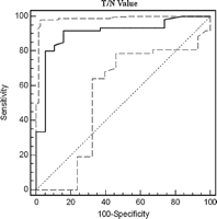
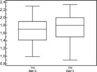
In patients with neoplastic lesion, the mean T/N ratio value ± SD was 1.72 ± 0.35 (0.9–2.34 range). On the other hand, in patients with benign lesions the mean T/N ratio value ± SD was 1.14 ± 0.25 (0.7–1.87 range). In 2/60 patients affected by malignant lesion T/N ratio was ≪1.14. In benign nodules, only one patient (tuberculoma) had T/N ratio value >1.72. The semiquantitative analysis showed a markedly higher T/N ratio in malignant lesions in respect of benign lesions (Student’s t-test, test statistic t: 6.722; degrees of freedom: 77; 95% CI: 0.417–0.769; p ≪ 0.0001). ROC curve (Fig. 1 ) shows that for a T/N value >1.23, the value of sensitivity, specificity, PPV, and PNV were 91%, 84%, 94%, and 76%, respectively (area under the ROC curve: 0.903; standard error: 0.034; 95% CI: 0.815–0.958). Further, there was no significant correlation between T/N ratio and the size of tumor (≪ or >3 cm in diameter) (Mann–Whitney test) (Fig. 2 ); age, sex, and histologic type of lung lesion did not affect the SPECT results (Fisher’s exact test).

ROC curve shows that for a T/N value higher than 1.23, sensitivity, specificity, PPV, and PNV is 91%, 84%, 94%, and 76%, respectively.

Regarding tumor size, there was no significant difference of T/N ratio in patients with malignant lesion ≪3 cm compared with those >3 cm in diameter ((Mann–Whitney test, p = 0.533).
In 57 patients the metastatic involvement of mediastinal lymph nodes was assessed by histologic examination after mediastinoscopy or thoracotomy. Metastatic mediastinal lymph nodes were found in 3/57 patients. In two of these patients 99mTC-MIBI SPECT (T/N > 1.89) was positive while CT was positive for suspected metastases in only one patient. None of the patients with negative lymph nodes had SPECT positive results. 99mTc-MIBI SPECT showed a specificity and PPV of 100% in the detection of mediastinal lymph nodes with sensitivity, and PNV of 66% and 97%, respectively. Age, sex, histologic type, and size of lymph node did not affect the SPECT results.
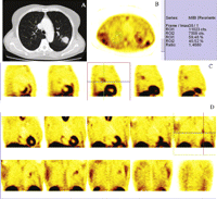
An illustrative image of 99mTc-MIBI SPECT in a patient with malignant lung lesion is shown in Fig. 3 .

A 55-year-old woman with squamous cell carcinoma. CT scan shows primary tumor of the left upper lobe surrounded by normal lung tissue, and not greater than 3.0 cm in its largest diameter without evidence of hilar or mediastinal adenopathy (Part A). 99mTc-MIBI SPECT in transaxial view reveals good visualization of cancer. Drawing regions of interest (ROIs) over the tumor uptake (T) and in the controlateral normal side (N) is performed to calculate T/N ratio value (Part B). 99mTc-MIBI SPECT in sagittal (Part C) and coronal (Part D) views demonstrate area of increased abnormal uptake corresponding to lung cancer.
4 Discussion
The best strategy for treating a patient with a lung lesion depends on the probability of cancer for that particular lesion. Conventional imaging methods, such as chest computed tomography and magnetic resonance, continue to play important roles in the detection of lung cancer and in tumor staging. Radiological findings suggestive of malignancy are thickness of the cavity wall and the presence of speculated or nodular edge, whereas central, laminated, or diffuse calcifications are more likely to be related to a benign etiology. However, despite the advances in morphological imaging, these modalities provide anatomic and morphologic information, but they often fail to distinguish between malignant and non-malignant lesions in the lung. For lesions with patterns of calcification that are radiologically ‘indeterminate’ and for noncalcified lesions, the differentiation between the malignant and benign usually requires a more invasive approach. Invasive procedures are often undertaken to obtain a specific tissue diagnosis and the presence of benign lesion in 60% of removed nodules indicates a need for an approach that better enables lung preservation [14]. Because the evaluation of such lung lesions represents a daily dilemma for surgeons, a non-invasive diagnostic method for the identification of malignancies is desirable. Therefore, the development of functional imaging examinations represents a significant advance in the diagnostic assessment of patients with suspected lung cancer. Despite the growing applications of PET and PET/CT, SPECT studies are still very useful in numerous conditions and single-photon radiopharmaceuticals have demonstrated clinical value in a wide variety of neoplasms, including lung cancer [15,16]. Since the late 1980s an increasing number of papers have appeared describing 99mTc-MIBI uptake in breast cancer, lymphoma, mediastinal and pulmonary metastases from thyroid cancer [12,13,17–20]. 99mTc-MIBI is a widely used myocardial perfusion imaging agent whose myocardial distribution is proportional to regional blood flow. The mechanism of 99mTc-MIBI uptake in tumor cells is not yet well known. However, in vitro studies have demonstrated that the tracer uptake, favored by an increased blood flow and capillary permeability and high metabolic activity of neoplastic cells, is strictly dependent on cell membrane and mitochondrial potentials. 99mTc-MIBI accumulates within mitochondria and cytoplasm of cells on the basis of electrical potentials generated across the membranes. Since malignant tumors maintain a more negative transmembrane potential due to their increased metabolic requirements, this has postulated to cause increased accumulation of MIBI in malignant mass [21,22]. In vivo studies have investigated the diagnostic value of 99mTc-MIBI to detect lung cancer. Hassan et al. [17] report data on 22 patients with primary lung cancer in which the sensitivity of planar 99mTc-MIBI is 95% (21/22). At semiquantitative evaluation, in patient with lung cancer the mean T/N ratio is 1.45 ± 0.32 (1.08–2.22) and it is significantly affected by type (in SCLC higher than NSCLC) and size of the tumor. Nosotti et al. [18] in studying 116 patients with suspected lung cancer, find specificity, sensitivity, PPV, NPV, and accuracy value of 100%, 89.8%, 100%, 62.9%, and 91.4%, respectively. In addition, the authors compare the accuracy of 99mTC-MIBI with that of CT in the staging of mediastinal lymph nodes and conclude that 99mTC-MIBI is more accurate than CT (p ≪ 0.011). Minai et al. [12] report on the use of 99mTC-MIBI in patients with radiological evidence of lung lesions, obtaining a sensitivity of 85.7% (18/21) for the detection of lung cancer, a specificity of 100%, a PPV of 100%, and a NPV of 57%. In contrast, Kao et al. [19] conclude that 99mTc-MIBI is of limited use in the differentiation of lung lesion. The authors report that in 54 patients with single solid lung masses, 99mTC-MIBI detects 65% (30/46) of the lung malignancies and 75% (6/8) of the benign lesions. The diagnostic sensitivity, specificity, and accuracy in differentiating malignant and benign lesions are 65%, 57%, and 70%, respectively. On the basis of these preliminary but encouraging findings, we investigate the role of 99mTC-MIBI SPECT to better evaluate its diagnostic accuracy. We study a total of 79 patients (60 with malignancies and 19 with benign lung lesions) and our data confirm that 99mTC-MIBI is a good radiotracer to detect lung malignancy. This technique identifies 91.6% (55/60) of the lung malignancies and 73.6% (14/19) of the benign lesions. Sensitivity, specificity, PPV, and PNV of SPECT results by quantitative analysis are 91%, 73%, 91%, and 73%, respectively. Compared to the results reported by Nosotti et al. [18] and Minai et al. [12], the sensitivity of our data is similar but specificity is lower. We suspect that the somewhat reduced specificity seen in our group is related to misinterpreted areas of radiotracer accumulation by visual evaluation. If the activity in the lesion is greater than the activity seen in the normal lung, there is not the possibility of disagreement, consequently SPECT images are considered as ‘positive’ for malignancy. Differences of opinion are possible when an increase of radiotracer is low or in the presence of lung lesions characterized by a focal uptake with a central hypoactive focus as well as a ring-like appearance of increased uptake. In these case interobserver variability is extremely high, and it should not be resolved by a consensus of readers. In 3/5 patients with benign lesion, the increase of tracer is low but SPECT images are considered as ‘positive’. In 2/5 patients affected by malignant lesion with central necrotic area, the uptake of tracer is localized only in the periphery of the lesion with a central hypoactive focus. In both cases, SPECT images are considered as ‘negative’. Thus, we analyze the SPECT result by semiquantitative analysis according the value of T/N ratio for avoiding the possible inaccuracy of visual evaluation. In case of equivocal lesion, semiquantitative evaluation gives additional information for a differential diagnosis between malignant and benign lesions. The T/N ratio values in malignant lesions (mean value 1.72) are higher compared with those of benign lesions (mean value 1.14) with significant statistical differences (Student’s t-test, p ≪ 0.0001). In addition, ROC curve shows that for a T/N value higher than 1.23, sensitivity, specificity, PPV, and PNV is 91%, 84%, 94%, and 76%, respectively. This indicates that if the T/N ratio value of lung lesion is higher than 1.23 values, the patient is very likely to have a malignant lesion. Consequently, the clinical implication is that, in the case of a pulmonary lesion whose nature is indeterminate after non-invasive evaluation, a positive 99mTC-MIBI SPECT may help to avoid unnecessary invasive diagnostic testing and allow the physician to proceed directly to thoracotomy. In addition, it may be helpful in cases where non-invasive testing is suspicious for malignancy but the patient has impaired cardiopulmonary function such that the risks of thoracotomy are judged to be high. The findings of a high specificity and PPV of our study may depend on the selection of the patients, namely, on the high prevalence of malignant lesions. In our study group, the vast majority of the lesions are malignant (60/79), which is in contrast to most series reporting on solitary pulmonary nodules [23]. However, several reasons may affect the low percentage of benign lesion observed. First, the percentage of malignant lesions can significantly depend on the age and smoking habits. For example, lung carcinoma is extremely rare in patients less than 30 years of age whereas in patients aged more than 50 years, over one half of lung nodule are malignant lesions; in our study, the mean age of patients are 66 years and 91% of them are smokers. Second, all patients of our study group are referred from a thoracic surgery unit with prevalence of cancer probably higher than that expected if patients are referred from a pulmonary medicine clinic. Third, the high prevalence (75.9%) of malignant lesions might be related to selection bias as we include the patients (50/79) with a nodule diameter over 3 cm. However, with the exception of size >3 cm in diameter, the lesions of our study group are ‘indeterminate lesions’ because they are noncalcified and no specific diagnosis could be achieved on the basis of morphological imaging. In addition, it is not unexpected that in our population neoplastic lesions tend to be larger than the benign lesions (mean diameter, 3.9 cm vs 3.3 cm, respectively) but it does not mean that a correlation exists between quantitative uptake of 99mTC-MIBI SPECT and the diameter of nodule or that a larger nodule will be easier to identify on MIBI SPECT. Of the five malignant nodules with T/N ratio value less than 1.23 value, only one nodule has a diameter less than 3 cm. Thus, the small size of the lesion does not represent the most important factor responsible for this negative case in which other factors should also be taken into account, such as metabolic and/or biological factors [21,22], not evaluated in these patients. Moreover, 99mTc-MIBI SPECT demonstrates similar accuracy in evaluation of nodules that are ≪ or >3 cm in diameter (Fig. 2); thus tumor detectability is independent of the tumor size. Our data are in contrast to the findings of Hassan et al. [17] and Minai et al. [12] who report a significant relationship between uptake of MIBI and nodule size but no cut-off size is identified by authors. On the other hand, in concordance with our findings, Nosotti et al. [18] report that no correlation is found between the size of nodule and the 99mTC-MIBI SPECT results.
In addition, in our series of malignant lesions we do not observe any significant correlation between MIBI uptake and histological type of the malignant lesion; of the five malignant nodules with T/N ratio less than 1.23, two are squamous cell carcinoma, one is adenocarcinoma, one is large cell carcinoma, and one is a metastatic lesion. Our data are in concordance with the findings of Nosotti et al. [18] but in contrast with other reports [12,20]. Nishiyama et al. [20] find that squamous cell carcinoma has lower T/N ratios than adenocarcinoma and small cell carcinoma. Hassan et al. [17] confirm that MIBI uptake is higher in SCLC than NSCLC and the authors highlight that undifferentiated squamous cell carcinoma can be negative by MIBI SPECT scanning. However, we do not have a definitive conclusion about correlation between histological type and SPECT results for several limitations in terms of the patient population. First, in our series only 1/60 patient (1.6%) with lung malignancy is affected by SCLC with respect to 27.2% of cases reported by Hassan [12]. Second, the vast majority of our malignant lesions are squamous cell carcinoma, with low prevalence of adenocarcinoma and of large cell carcinoma.
Regarding the value of sensitivity and NPV of our report, 3/19 (15.7%) benign nodules have a T/N ratio value over 1.23 with sensitivity of 91% and PNV of 76%. The clinical implication is that, if the lung mass has a T/N ratio value less 1.23, in patients with a lesion that is estimated to have a low likelihood of being malignant (age ≪30 years, no history of previous malignancy, no smoking history, no features of malignancy at radiological evaluation) lung cancer can be ruled out with a reasonable degree of reliability and invasive diagnostic procedures like thoracotomy may be avoided with the potential for significant cost savings.
Finally in lymph node surgical staging, 3/57 patients have lymph node metastasis, 2 have positive value of SPECT, and 1 of these have CT suspected metastases (>1 cm). None of the patients with negative lymph nodes has SPECT positive results. Sensitivity, specificity, PPV, and NPV of SPECT results are 66.7%, 100%, 100%, and 97.9%, respectively. According to the findings of Nosotti et al. [18], we believe that in preoperative staging of lung cancer, this technique could play a clinical role in reducing the number of invasive procedures such as mediastinoscopy and mediastinotomy in selected patients, particularly in those with enlarged lymph node at CT and positive to 99mTC-MIBI SPECT. However, because of the small number of patients, more data are needed from larger series to develop decision-making protocols based upon MIBI results for mediastinal staging in patients with proved lung cancer. Finally, a further interesting point of discussion is a comparison of PET and 99mTC-MIBI SPECT results to estimate the value of these modalities in differentiating malignant and benign pulmonary lesion. However, PET is not available at our hospital and few patients of our study group have performed PET at other sites. Consequently, we are not able to execute this relationship at the moment but we hope that it will be the aim of the next paper.
5 Conclusion
Our results seem to confirm that 99mTc-MIBI SPECT is a reliable diagnostic tool in the finding of lung cancer particularly in cases in which radiological evaluation is indeterminate. This procedure is readily available, safe, and more cost effective in comparison with FDG PET. In our country, the cost of whole-body FDG PET is Euro 950.00, whereas a 99mTc-MIBI SPECT costs Euro 100.00. Compared to visual analysis, semiquantitative study is more accurate and gives additional information for a differential diagnosis between malignant and benign lesions; it facilitates the making of a decision as to positive or negative scintigraphic character of the equivocal lesions. Finally, because of the small number of patients further studies are required to corroborate the clinical role of 99mTc-MIBI SPECT in the evaluation of suspected lung cancer and its specificity.
References
Author notes
Presented at the 16th European Conference on General Thoracic Surgery, Bologna, Italy, June 8–11, 2008.
- radionuclide imaging
- technetium tc 99m sestamibi
- technetium 99m
- lung
- single photon emission computed tomography
- small cell carcinoma of lung
- squamous cell carcinoma
- adenocarcinoma
- carcinoma, large cell
- neoplasm metastasis
- photons
- roc curve
- diagnosis
- neoplasms
- chest
- lung cancer
- clinical diagnostic instrument
- mediastinal lymph node group


