


Abstract
Background: Several methods have been utilized to prevent pericardial and retrosternal adhesions, but none of them evaluated the mesothelial regenerative hypothesis. There are evidences that the mesothelial trauma reduces pericardial fibrinolytic capability and induces an adhesion process. Keratinocyte growth factor (KGF) has proven to improve mesothelial cells proliferation. This study investigated the influence of keratinocyte growth factor in reducing post-surgical adhesions. Methods: Twelve pigs were operated and an adhesion protocol was employed. Following a stratified randomization, the animals received a topical application of KGF or saline. At 8 weeks, intrapericardial adhesions were evaluated and a severity score was established. The time spent to dissect the adhesions and the amount of sharp dissection used, were recorded. Histological sections were stained with sirius red and morphometric analyses were assessed with a computer-assisted image analysis system. Results: The severity score was lower in the KGF group than in the control group (11.5 vs 17, p = 0.005). The dissection time was lower in the KGF group (9.2 ± 1.4 min vs 33.9 ± 9.2 min, p = 0.004) and presented a significant correlation with the severity score (r = 0.83, p = 0.001). A significantly less sharp dissection was also required in the KGF group. Also, adhesion area and adhesion collagen were significantly lower in the KGF group than in the control group. Conclusion: The stimulation of pericardial cells with KGF reduced the intensity of postoperative adhesions and facilitated the re-operation. This study suggests that the mesothelial regeneration is the new horizon in anti-adhesion therapies.
1 Introduction
Postoperative adhesions are formed after surgical procedures on heart and great vessels as part of the healing process. The scar tissue makes re-entry hazardous increasing the rate of iatrogenic lesions. Literature describes 4–9.1% [1,2] accidents during resternotomy and adhesiolysis where graft structures were the most damaged (46.4%) followed by the right ventricle (21.4%) [2].
As re-operations account for 8.5–34% of heart surgeries nowadays [3–6], various methods have been investigated to prevent or decrease the severity of the postoperative adhesions. Heterologous pericardium [7], anti-inflammatory drugs [8], synthetic rubbers [9], fibrinolytic agents [10] and biopolymers [11,12] are examples of therapies explored for such purpose.
Since 1971, it is known that mesothelial cells are responsible for fibrinolytic properties of the coelomic cavities [13] and the injury to these cells are the trigger for adhesion formation [14]. In 2000, Adamson demonstrated that the proliferation of mesothelial cells can be up-regulated by the keratinocyte growth factor (KGF) [15,16]. In theory, this growth factor could hasten the pericardium recovery, which could improve the fibrinolytic capability, thus reducing the adhesion formation.
Therefore, the aim of this study was to explore a new field in postoperative adhesion prevention; the regenerative method. The research hypothesis was to evaluate the influence of the use of recombinant human KGF (r-KGF) in the reduction of post-surgical pericardial adhesion.
2 Material and methods
Twelve male Large White swine, weighing from 15 to 20 kg, were used in this research. The Heart Institute (InCor), University of São Paulo ethics committee, approved the study protocol. All animals received human care in compliance with the ‘Guide for Care and Use of Laboratory Animals’ published by the National Institutes of Health (NIH publication 85-23, revised 1996) and the ethics principles in animal research established by the Brazilian College of Animals Research (COBEA) were followed.
2.1 Preparation of experiment samples
The r-KGF was purchased in a highly purified state (≥95%) from Sigma (Sr. Louis, MO, cat #: K 1757, Lot #: 75K1562). Stock solutions were made after an initial dilution with a phosphate-buffer saline filtered through a 0.2 μ membrane. Aliquots were stored at −20 °C at 111 ηg/ml concentration.
At the time of use, the aliquot were thawed and diluted in 12 ml sterile bidistilled water. The KGF group sample consisted of a 15 ml solution containing 22 ng/ml r-KGF. The same volume of 0.9% (w/v) sterile saline was used as control group sample.
2.2 Surgical protocol
The animals were fasted 12 h prior to the surgery. Anesthesia was induced with an intramuscular injection of ketamine hydrochloride (10 mg/kg) and atropine (0.05 mg/kg). A prophylactic dose of penicillin-streptomycin veterinary antibiotic (Pentacilin C®, Fort Dodge, São Paulo, Brazil) was administered. A venous line was established in the ear and a saline solution was infused at a rate of 3 ml/kg/h to replace the fasting and non-sensed water losses. All had continuous two-lead electrocardiograph monitoring during the operative procedure. Endotracheal intubation was performed after a venous bolus of thiopental sodium (10 mg/kg) and phentanyl (0.05 μg/kg). Artificial respiration was obtained with a volume-control ventilator (Antares®, Calgimed, São Paulo, Brazil). Anesthesia was maintained with 0.5–2% isoflurane.
All surgical steps of the adhesion induction protocol (abrasion, blood instillation and desiccation) were performed by the same surgical team in a blinded manner. After standard skin preparation, a 5 cm thoracotomy was performed through the fifth right intercostal space. A pericardiotomy, anterior to the phrenic nerve, was made and the heart exposure was improved with traction ligatures at the pericardium edges. The epicardium and parietal pericardium related to right ventricle atrium, right and left ventricle were abraded with a 1.5 cm × 1 cm sandpaper (Adalox® T 223, Norton Abrasivos, São Paulo, Brazil) in 10 manual movements. Polyester 2.0 (Mersilene®, Ethicon, São Paulo, Brazil) sutures were performed in the aorta and right atrium and these were tied in a way to simulate the cardiopulmonary bypass cannulation. The pericardial cavity was covered with 20 ml of autologous blood and 30 min delay occurred until the clots had been aspirated. A fenestrated catheter was inserted through a small pericardial orifice and the cavity was closed with a running suture.
At this point, the surgical team was informed about the animal randomization and the KGF sample (KGF group) or saline (control group) was injected into the cavity through the intrapericardial catheter. The catheter was removed and the orifice tied. A thoracic drain was placed to evacuate the air in pleural cavity, and the chest was closed in three layers. The anesthetic gas was suspended and the animals allowed to awake. The postoperative analgesia was obtained with intramuscular doses of morphine sulphate (0.2 mg/kg).
The animals were observed three times daily for the first 3 days and remained isolated until the 5th postoperative day.
2.3 Re-operation
Eight weeks after the initial procedure, following the same anesthesia protocol, the animals were submitted to a median sternotomy by a blinded surgeon. Adhesion formation was evaluated in six intrapericardial areas: in anterior, lateral and inferior heart surfaces, in the right atrial suture, in the aortic suture and in the pericardial suture line. An observer, also group-blinded, graded these areas using a scale system: grade 0, indicating that adhesions did not exist; grade 1, adhesions were filmy, light, with a foamy dissection plane; grade 2, where adhesions were intermediate requiring some sharp dissection but most of them were lysed by digital manipulation; grade 3, adhesions were dense, easily bleeding adhesions, with marked obliteration of tissue planes and required exclusive sharp dissection.
The adhesion score was defined as the sum of the adhesion grades in examined areas. One score was established for each animal.
The dissection time was measured with a digital chronometer. This interval calculated the time spent since the pericardium opening until the end of adhesiolysis in all six predefined areas. Also, the re-operation was recorded continuously, in two angles, by two digital cameras (Finepix S9600, Fuji, Japan) to quantify how many times it was necessary to use a sharp dissection. After the procedure, the images were reviewed and the scissors cutting movements were counted. The average of sharp movements registered by two cameras was defined as the sharp dissection variable.
At the end of the procedure the animals were euthanized with an overdose of thiopental sodium and a bolus of 19.1% (w/v) potassium chloride.
An in-block fragment was obtained from the mid-distance between the superior and inferior vena cava and was immersed in 10% (w/v) paraformaldehyde. This tissue section, from inside to outside, was formed by the atrial wall, the adhesion tissue and parietal pericardium.
2.4 Light microscopic examination
The fixed histological specimens were embedded in paraffin, sectioned into a 5 μm and stained with sirius red [17]. Sections were examined by light microscopy using a 5× magnification objective lens. Images were digitalized using a digital video camera (JVC KY-F55B, Japan) with a resolution of 768 × 576 (vertical × horizontal) pixels. Pixel size was converted into micrometers and the image analysis was performed using image analysis software (Leica Quantimet Q500MC, Cambridge, UK).
The morphometric evaluation consisted in measures of pericardium, adhesion and epicardium areas and in semi-quantitative analyses of collagen content of these areas.
2.5 Statistical analysis
Categorical variables are listed as median (max–min) and continuous are listed as average ± standard deviation. Data analysis was performed by GraphPad Prism, version 5.01. The adhesion score, the dissection time, the sharp dissection and histological parameters were evaluated by Mann–Whitney test. The Spearman test was used for non-parametric correlation analysis. Linear and non-linear regression analyses were applied to evaluate the relation between the adhesion score and the dissection time and the amount of sharp dissection. Statistical significance was achieved at p ≪ 0.05.
3 Results
There were no postoperative deaths in either group. In the KGF group, one animal suffered a skin and subcutaneous suture dehiscence that was treated locally. In the control group, one animal had a right ventricle lesion caused by the bone saw during the sternotomy and died before the end of the procedure.
3.1 Macroscopic findings
The results of the adhesion intensity in all studied areas are shown in Table 1 . KGF reduced the adhesion scores when compared with the controlgroup (p = 0.005). Score, expressed as median (min–max), were 17.0 (15.0–18.0) in the control group and 11.5 (9.0–12.0) in the KGF group (Fig. 1 ). All areas evaluated had a significantly lesser adhesion severity score in the KGF group, except by the pericardium suture line.
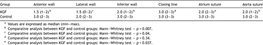
Macroscopic evaluation of adhesion severity in each analysed segmenta.
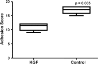
Adhesion score at 8th postoperative week in the two groups.
In the control group, the mean time required to free the heart was 33.9 ± 9.2 min. In the KGF group, the time was significantly reduced to 9.2 ± 1.4 min (p = 0.004) (Fig. 2 ).
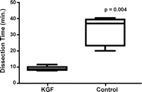
Differences in dissection time spent to free the heart and great vessel of adhesions in the two groups.
The amount of sharp dissection employed in adhesiolysis was significantly less in the KGF group (81 ± 28). In contrast, 291 ± 101 sharp movements were necessary in the control group (p = 0.004) (Fig. 3 ).
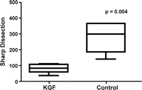
Requirement of scissor movements to carry on with the adhesiolysis in the two groups.
There were significant correlations between the adhesion score and the dissection time (r = 0.83, 95%CI 0.45–0.95) and between the adhesion score and the sharp dissection required to free the heart (r = 0.96, 95%CI 0.84–0.99). The regression analysis identified a significant relationship between the adhesion score and the sharp dissection required (Fig. 4 ), which presented an exponential behavior. The same observation was shown between the adhesion score and the dissection time (Fig. 5 ).
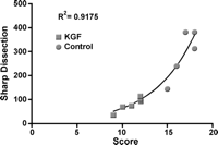
Exponential relationship between the adhesion score and the amount of sharp dissection performed for adhesiolysis. It should be observed that this data could not be acquired in one animal due to a re-entry accident.
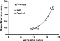
Exponential relationship between the adhesion score and the time used for dissection. It should be observed that this data could not be acquired in one animal due to a re-entry accident.
3.2 Microscopic findings (Table 2)
The epicardium, the adhesion and the parietal pericardium areas were more prominent in the control group than in the KGF group (p = 0.02, p = 0.009, p = 0.04; respectively). Also, the sirius red stain (Fig. 6 ) demonstrated that the collagen’s measured area was increased in the control group when compared with the KGF group, in the epicardium, in the adhesion and in the parietal pericardium (p = 0.04, p = 0.03, p = 0.04; respectively). The proportion of collagen into the scar tissue was not different at the three areas analysed.
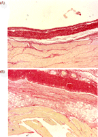
Sirius red staining. (A) KGF group with a minimal epicardial reaction, a scant non-compacted fibrous tissue creating the adhesion and a usual parietal pericardium with a standard pleural interface (×5). (B) Control group showing a epicardial reaction, the adhesion formed by a dense non-compacted fibrous tissue and a thickened parietal pericardium with a broad interface with the parietal pleura (×5).
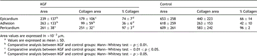
Morphometric evaluation of the adhesion fragmenta.
4 Discussion
This study showed, for the first time, that the use of a growth factor targeted toward mesothelial cells proliferation/regeneration is able to reduce the severity of adhesions and re-operation challenges, as shown by the reduction in the time required to free the heart from the other surrounding structures using less sharp dissection that, in a clinical setting, could have an impact in the morbidity rates.
The barrier methods using bio-absorbable biopolymers released in a gel composition or in a sheet layer showed good results [11,12]. These methods are in accordance with the mesothelial healing process [14] and permit that visceral and parietal serosas both become disclosed, while the mesothelial regeneration proceeds.
Normally, the mesothelial cell repair begins within the first day of injury and in 8–10 days it is finished. Mesothelial cells are responsible for pericardial fibrinolysis [14] and it is known that operative trauma decreases this activity [18]. Surgical aggression halts the local fibrin degradation process, which permits the formation of fibrin bands that will be transformed in scar tissue. Two ways to restore fibrinolytic capabilities of the serosal membranes are to supply a fibrinolytic agent [10] or to transplant the source of the fibrinolytic agent; mesothelial cells [19]. Both have been suggested as strategies to reduce peritoneal adhesions, but unfortunately the direct application of fibrinolytic enzymes after heart surgeries is unsafe and the problem of a cell source is critical because procedures to obtain mesothelial cells implicate a prior extraction and cell culture. Nevertheless, through mesothelial regeneration the pericardium fibrinolytic function can be restored [20] and the faster this can be accomplished the less fibrin will remain to be substituted by collagen.
Keratinocyte growth factor, also known as fibroblast growth factor-7, belongs to the heparin-binding growth factors family. Their potent mitogenic activity on epithelial cells is exhaustively known, but only in 2000 was it discovered that this growth factor can increase mesothelial cell proliferation [15], thus, opening the possibility to a faster mesothelial repair restoring pericardial fibrinolytic characteristics.
The injury to the mesothelial cells is the trigger to the adhesion formation process. The present study explores a new method of preventing the adhesions that is based on the human recombinant FGF-7 targeted to the mesothelial cells proliferation. Despite concerns about the use of human growth factor in others mammal species, human KGF effects in a porcine model have been demonstrated already [21]. Another point involved in KGF therapeutic use is their potential for cancer development or stimulation. Neither KGF-1/FGF-7 nor, their homolog [22], KGF-2/FGF-10 has been implicated in tumorigenesis, in enhanced tumor growth or in the inhibition of the cytotoxicity of cancer treatments. Although some data suggest that KGF might be beneficial in some tumors like prostate, bladder and salivary, special caution should be taken when using this growth factor in patients with breast and stomach cancers due to pending information about its activity in these contexts [23].
A recent report [24], also exploring the concepts of the mesothelial regeneration by cellular proliferation stimuli, could demonstrate a reduction in the adhesion formation using an angiogenic agent (ginsenoside) extracted from Panax ginseng to improve the neovascularization of an acellular pericardium. Although not using a direct mesothelial proliferative agent, this study also demonstrates that the studies in postoperative adhesion prevention should focus on mesothelial regeneration.
In addition, to reduce the postoperative pericardial adhesions this work introduces the concept that the time spent in dissection and the amount of sharp instrument use can describe accurately the severity of the adhesions. Also, this model performed a discrete modification in the abrasion/blood/desiccation, where the gauze used in abrasion was replaced by sandpaper. This alteration created a uniform injury and induced consistent and homogeneous formation of adhesions in the control group allowing a clear clarification of KGF effects in the test group.
The microscopic study shows that the animals of the KGF group had a lesser area of adhesion and collagen, but the proportion of collagen in the scar tissue did not differ from the control group, which suggested that instead of a modification in the healing process, a reduction occurred in the extension of the fibrin matrix that would be replaced by collagen. The possible reduction of the fibrin matrix corroborates the hypothesis that the occurrence of less pericardial adhesions with the use of KGF is based on the resumption of the pericardial fibrinolytic function by mesothelial cells.
Although the effectiveness of the KGF observed here opened a new field in the adhesion prevention research, the closed pericardium model employed in this study brought a practical concern. In the first instance, this could be thought of as a limitation because most heart surgery usually uses postoperative drains. As KGF has ionic attraction to collagen [25] and this molecule is exposed in a mesothelial denuded area, it is not possible to predict if the use of postoperative drains could have influence in its effectiveness. Furthermore, many surgeons do not routinely close the pericardium and the KGF effect for the unclosed pericardium is unknown. Nevertheless, others studies should answer these queries and, also, they should explore the use of the KGF combined with biopolymers gels and biopolymers sheets.
In conclusion, the regenerative method is a new and efficacious therapy to be employed in anti-adhesion treatment area. The results of this study show that KGF reduces the severity of the adhesions and can reduce surgical time and the use of sharp dissection, but new studies should be developed to directly measure the influence of KGF in the pericardial fibrinolytic activity, and in the accelerated healing of the pericardial defects by the new mesothelium.
Acknowledgments
We thank the assistance of Mr Pedro Castro and Mrs Flavia Luana and the non-financial support of the Marcio Cunha Hospital and of the Faculdade de Medicina do Vale do Aco.
References
Author notes
The authors disclose any commercial associations that might pose a conflict of interest in connection with the submitted article. This study is subject of a patent-licensing arrangement.
