


Abstract
Objective: In the study, we made the pharyngoesophageal functional assessment and menometric study on the two kinds of anastomosis (traditional hand-sewn anastomosis and side-to-side stapled anastomosis) for the further evaluation and application of cervical esophagogastrostomy. Patients: The study included 17 patients with esophageal squamous cancer from March 2006 to May 2008. Eight patients had undergone total esophagectomy and traditional hand-sewn technique in CEGA. The other nine patients had undergone total esophagectomy and side-to-side stapled technique in CEGA. All the 17 patients were studied for 3 months after the operations. The complete data, such as esophagogastroscopy, barium swallow and manometric studies, were obtained for each participating patient. Results: In the hand-sewn group of eight patients, four patients (50%) reported clinical significant symptoms of cervical dysphagia. Two patients (11.1%) reported clinical significant symptoms of cervical dysphagia in the side-to-side group of nine patients. There is a statistically significant difference between the hand-sewn group of patients (n = 8) and the side-to-side group of patients (n = 9) with respect to overall mean anastomotic diameters (1.688 ± 0.26 cm vs 3.012 ± 0.17 cm, p = 2.10 × 10⁻⁸). In the eight patients who underwent hand-sewn technique, there were four symptomatic patients with poor menometric datum, such as anastomotic hypertensive peristaltic activity, confusing inversion of anastomotic and midcervical esophageal pressure, and consequently poor compliance of the pharyngoesophageal segment (pharyngeal shoulder pressure). By contrast, there was only one symptomatic patient with poor menometric data in the nine patients who underwent side-to-side technique. Conclusion: The side-to-side stapled technique is conducive to decrease complications of postoperative dysphagia and is helpful for improving pharyngesophageal and anastomotic menometric function. The anastomotic technique deserves more attention and further applications.
1 Introduction
According to the statistics from WHO, the mortality of esophageal cancer in China is the highest in the world [1,2]. Chinese surgeons are making efforts to improve the techniques in the operation of esophageal cancer resection and esophagogastric anastomisis. Nowadays cervical esophagogastric anastomosis (CEGA) is a widely accepted procedure because of larger resection margins and less dangerous anastomosis leakage [3,4]. As a result, the technique of hand-sewn anastomosis in cervical esophagogastrostomy is becoming very common due to better tumor eradication, and reduced morbidity and mortality associated with anastomotic breakdown. However, traditional hand-sewn anastomosis in cervical esophagogastrostomy brings high risks of leakage, stricture, and pharyngoesophageal dysphagia [5,6]. In 2000, Orringer et al. [7] introduced a very novel CEGA technique incorporating both the hand-sewn technique and the using of endo-GIA stapler devices. That procedure is called side-to-side stapled anastomosis and is allegedly associated with a lower rate of anastomotic complications compared to the traditional hand-sewn anastomosis in several recently published reports [8,9]. As a result, this technique attracts the interests of more and more surgeons. However, all the published papers on side-to-side stapled anastomosis have focused on clinical observation and there is not any control study on functional and menometric study of side-to-side stapled anastomosis and traditional hand-sewn anastomosis. In the study, we made the pharyngoesophageal and anastomotic functional assessment regarding the two kinds of anastomosis for the further improvement and application of cervical esophagogastrostomy.
2 Patients and method
The study protocol was reviewed and approved by the research ethics board in Daping hospital (TMMU-DPH/2008-012), and informed consent was obtained from all patients who agreed to participate in the study.
2.1 Patients
From March 2006 to May 2008, the study included 17 patients with esophageal squamous cancer. Eight patients (six males, two females; median age 65 years; five patients with midthoracic esophageal cancer and three patients with upper thoracic esophageal cancer) had undergone total esophagectomy and traditional hand-sewn technique in CEGA. All these eight patients were included in the ‘hand-sewn group’. The other nine patients (five males, four females; median age 63 years; six patients with midthoracic esophageal cancer and three patients with upper thoracic esophageal cancer) had undergone total esophagectomy and side-to-side stapled technique in CEGA. The nine patients were included in the ‘side-to-side stapled group’. All 17 patients were studied in the 3 months after the operations. Complete data were obtained for each participating patient. No patient had a postoperative anastomotic stricture (early or late). All patients regularly attended 3-monthly postoperative follow-up clinics where a physical examination and chest radiography was performed. No patient had radiographic evidence of recurrent or metastatic tumor, and all participating patients were considered disease-free.
2.2 Operative techniques
The operative technique consisted of a total thoracic esophagectomy with mediastinal lymphadenectomy via a right thoracotomy and gastric tube construction via a midline laparotomy incision with preservation of the right gastroepiploic artery [2]. Celiac lymph node dissection was also conducted during the laparotomy phase. Radical sweep of cervical lymph nodes was conducted and resection of cervical esophagus as far as possible from the superior margin of esophageal cancer was performed. Then the gastric tube was pulled up through the posterior mediastinum to the right cervical area for side-to-side stapled or hand-sewn CEGA. (1) The side-to-side stapled technique refers to the method of Orringer et al. [10] who performed the technique with an endo-GIA stapler on the anterior stomach wall. The endo-GIA II 3.5 mm stapler (Autosuture®, Norwalk, CT, U.S.A.) was used in the first step of the CEGA. The second phase of the CEGA, the anterior closure of the anastomosis site contained within the hood of the overlying esophagus, was performed by a hand-sewn method using Vicryl 5-0 (Ethicon®, Piscateway, NJ, U.S.A.) in interrupted sutures. (2) All hand-sewn anastomosis were constructed with a traditional, interrupted suture technique with the unabsorbable silk suture 4-0 (Xuelian®, Changhai, Nanchang, China). The posterior and anterior layer of the anastomosis took full thickness of both the esophagus and conduit. The posterior and anterior embedding of the anastomosis took between gastric placenta percreta and esophageal adventitia using the unabsorbable silk suture 4-0 (Xuelian®, Changhai, Nanchang, China). Each bite of the sutures was evenly placed (approximately 5 mm apart at a 5 mm depth) without pulling the sutures too tight to avoid strangulation of tissue.
Gastric drainage procedures were performed in all patients. Patients usually began taking fluids orally by the seventh day after surgery. To reduce the experimental deviation, all these operations were performed by a surgeon.
2.3 Clinical and objective studies
At the time of routine follow-up, all participating patients were evaluated clinically by an unbiased observer. In symptomatic patients, dysphagia to solids and liquids was further evaluated using a simple subjective dysphagia scale (0, no dysphagia; 1, difficulty with solids; 2, difficulty with semi-solids; 3, difficulty with liquids; 4, unable to swallow).
2.4 Esophagogastroscopy
Flexible esophagogastroscopy was performed under general anesthesia to exclude an anatomic obstruction, specifically an anastomotic stricture or recurrent tumor. The location of anastomosis was verified by esophagogastroscopy. The stapler, suture silk and borderline between esophageal mucosa and gastric mucosa could be conducive to the verification of anastomosis. The distance between incisor tooth and anastomosis should be recorded. In addition, the postoperative diameter of anastomosis was calculated in esophagogastroscopy as follows: (a) esophagogastroscopic camera faced the anastomosis as vertically as possible. (b) The distance between camera and anastomosis was fixed to be 1 cm with the aid of scaled steel-wire. (c) The pictures were taken consecutively and diameters of anastomosis were measured accurately with computer-assisted analysis (Medviewer 2.0, Huahai, China). The following additional objective studies were performed by consultant physicians, blind to other data and results.
2.5 Barium swallow
Video-fluoroscopy was performed on all patients by an independent consultant radiologist. Barium was used to define the anatomy of the reconstructed foregut, and to exclude an anatomic obstruction. The process of swallowing was assessed by evaluating transit of the barium bolus, elevation of the soft palate and hyoid, pharyngoesophageal coordination, and the identification of aspiration.
2.6 Manometric studies
Esophageal motility was studied by standard, water perfusion, stationary manometry (Medtronic DPT-6000, Smith Medical, Sweden) with computer-assisted analysis (Polygram 2.0, Smith Medical, Sweden) of the tracings according to a previously published protocol [11]. Briefly, a station pull-through technique was applied, and measurements were made at the function assessment of pharynx, the upper esophageal sphincter (UES), the midcervical esophagus (between the UES and anastomosis), and anastomosis (the peristaltic pressure can be detected on the point according to the distance between incisor tooth and anastomosis, which is previously verified by esophagogastroscopy).
2.7 Data analysis
All the data are presented as ‘mean ± standard deviation’. Quantitative data between the groups (e.g. anastomotic diameter of hand-sewn group and side-to-side group) were performed using the unpaired Student’s t-test. All data entry and analysis were performed with SPSS 13.0 software (Apache Software Foundation, Chicago, IL). A p value of 0.05 was considered statistically significant.
3 Results
3.1 Clinical assessment of cervical dysphagia
The postoperative complications are shown in Table 1 . All the complications had been treated successfully.
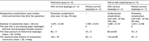
Postoperative complications, manometric data and diameter of anastomosis in hand-sewn group and side-to-side stapled group
In the hand-sewn group of eight patients, four patients (50%) reported clinical significant symptoms of cervical dysphagia. And in the side-to-side group of nine patients, two patients (11.1%) reported clinical significant symptoms of cervical dysphagia. However, the subjective severity of this symptom was variable, as alteration in patterns of swallowing was reported frequently, and was intermittent in frequency.
3.2 Anastomotic diameters
Objective endoscopic and radiologic studies did not demonstrate an anatomic obstruction. The anastomotic diameters of the two groups asymptomatically or symptomatically are shown in Table 1. Intriguingly, there was no statistically significant difference between asymptomatic patients and symptomatic patients with respect to mean anastomotic diameter in either hand-sewn group or side-to-side group. However, there was a statistically significant difference between hand-sewn group of patients (n = 8) and side-to-side group of patients (n = 9) with respect to overall mean anastomotic diameters (1.688 ± 0.26 cm vs 3.012 ± 0.17 cm, p = 2.10 × 10⁻⁸).
3.3 Manometric study
The interesting manometric characteristics of all the 17 patients are shown in Table 1.
Pharyngesophageal manometric tracing were performed successfully in all the 17 patients. A non-relaxing upper esophageal sphincter and a pharyngeal shoulder pressure are evident in the four symptomatic patients of hand-sewn group (Fig. 1a ). Besides, an anastomotic contractive high pressure appeared in all the symptomatic four patients of the hand-sewn group. And there is a statistically significant difference between the four symptomatic patients and the four asymptomatic patients with respect to the maximum peak pressure of anastomotic contraction (140 ± 18 mmHg vs 85 ± 5 mmHg, p = 0.0017). The mean pressure of midcervical esophagus is 75 ± 5 mmHg. There is not any statistically significant difference between the four symptomatic patients and the four asymptomatic patients with respect to the mean pressure of midcervical esophagus (80 ± 5 mmHg vs 75 ± 5 mmHg, p = 0.84).
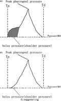
(a) Schematic diagram of a typical pharyngeal pressure tracing in the symptomatic patient of traditional hand-sewn group. Ta represents arrival of the bolus head; Td: completion of the pharyngeal-stripping wave. (b) Schematic diagram of a typical pharyngeal pressure tracing in the asymptomatic patient of side-to-side stapled group. Ta represents arrival of the bolus head; Td: completion of the pharyngeal-stripping wave.
In the two symptomatic patients of side-to-side group, there was one patient (maximum anastomotic contractive pressure = 110 mmHg) who had a non-relaxing upper esophageal sphincter and a pharyngeal shoulder pressure in the pharyngoesophageal manometric tracing, while the other one (maximum anastomotic contractive pressure= 80 mmHg) did not have any abnormality in upper esophageal sphincter or pharyngeal portion. And in the seven asymptomatic patients, pharyngeal shoulder pressure disappeared completely (Fig. 1b) and the upper esophageal sphincter relaxed completely. Obviously there was a statistically significant difference between hand-sewn group (n = 8) and side-to-side group (n = 9) with respect to the maximum peak pressure of anastomotic contraction (112 ± 32 mmHg vs 56 ± 24 mmHg, p = 0.0004). The mean pressure of midcervical esophagus is 70 ± 5 mmHg.
4 Discussion
CEGA via traditional hand-sewn technique is widely used for the treatment of esophageal carcinoma because of some outstanding benefits such as larger resection margins and less dangerous anastomosis leakage. However, as a coin has two sides, CEGA with traditional hand-sewn also has some disturbing complications such as high morbidity of postoperative anastomotic leak and anastomotic cicatrical stricture. Besides, altered swallowing after esophageal resection and reconstruction is reported frequently and may develop at any stage postoperatively, impacting significantly on quality of life [12]. And after the anatomic obstruction and recurrent obstructing tumor is excluded, the functional etiological reason should be considered in the cases with cloudy cervical dysphagia. A very popular cervical side-to-side stapled esophagogastric anastomosis appears to decrease the postoperative complications distinctly compared with traditional hand-sewn technique. For example, Blackmon et al. [13] reported that postoperative dysphagia was significantly higher in hand-sewn anastomosis at 56.5% vs 26.1% with side-to-side stapled (p = 0.04). And stricture requiring esophageal dilation was also increased in hand-sewn at 34.8% vs 8.7% with side-to-side stapled and 8.7% with circular-stapled (p = 0.04). However, very few studies have been performed for the contrasting evaluation of the menometric features in hand-sewn anastomosis and side-to-side stapled anastomosis. We designed the novel clinical experiment to evaluate the functional and menometric features of hand-sewn and side-to-side stapled technique. As an added incentive, we aimed to reveal the unclear mechanism of postoperative cervical dysphagia.
In our study, none of the patients had recurrent tumor; however, the clinical symptoms of dysphagia with various extent appeared in four patients of hand-sewn group and two patients of side-to-side group. The ratio of dysphagic risk came up to 50% in the eight patients with traditional hand-sewn technique, and it decreased to 11.1% in the nine patients with side-to-side stapled technique, which is consistent with the published report [13].
Pharyngeal swallowing is a mechanical process. The anastomotic hypertensive peristaltic activity (defined as mean esophageal pressures above 100 mmHg) appeared in the five of all the six symptomatic patients. One would conventionally expect a higher midcervical pressure as a result of distal obstruction if the anastomosis was causing an obstruction. However, the mean pressure in midcervical esophagus is 70–80 mmHg and yet the maximum peak pressure of anastomotic contraction is anything between 40 and 140 mmHg. The distance between anastomosis and midcervical esophagus is always less than 5 cm and the intervals between the detective roles in manometry are 5 cm. Hence, we cannot simultaneously screen the pressure and peristalsis in anastomosis and midcervical esophagus. We hypothesize that midcervical esophagus performs peristalsis to help the bolus to pass through anastomosis. However, the operations on the cervical esophagus may impair the nerve dominating cervical esophageal function and lead to the menometric dysfunction of esophageal muscle. Hence, the midcervical esophagus performs peristalsis simultaneously with anastomosis as ‘spasm’. Besides the midcervical esophagus cannot produce higher pressure to overcome anastomotic hypertensive peristalsis and to ‘propel’ the bolus through cervical esophagus. This might be the main cause of postoperative cervical dysphagia. Side-to-side stapled techniques could reduce the anastomotic hypertensive peristalsis (the midcervical esophagus need not overcome anastomotic hypertensive peristalsis) and the morbidity of postoperative cervical dysphagia. It is amazing that the anastomotic hypertensive peristaltic activity slowly stepped down and inversion of anastomotic and midcervical esophageal pressure became normal in all these patients after 3 months, which will be summarized in our other paper. It is necessary to determine the novel mechanism as the next step in research.
We analyze the reasons of appearance of pharyngeal shoulder pressure as follows:
We performed CEGA with radical sweep of cervical lymph nodes, especially the lymph nodes near recurrent laryngeal nerve. The procedures may have impacts on the nerve dominating cricopharyngeus muscles. What is more, for the sake of prevention of tumor recurrence, we resected cervical esophagus and made CEGA as far as possible from the superior margin of esophageal tumor. As a result, the anastomosis was closed to cricopharyngeus muscles and the anastomotic changes may impact cricopharyngeus function significantly.
In postoperative patients with CEGA, pharyngeal swallowing requires the thyrohyoid muscle groups to elevate the larynx, the glossopharyngeal musculature to propel the bolus, the cricopharyngeal to relax, and the cervical esophageal muscle and anastomosis to be compliant. This equates mechanically to three primary forces: (a) traction force (Ftraction), due to the contractions of the thyrohyoid muscles, resulting in the anterior-superior movement of the hyoid bone and, in turn, elevation of the larynx. (b) a bolus force (Fbolus) generated by the cricopharyngeus muscles, which propel the bolus into the pharyngoesophageal segment, and (c) inversion of anastomotic and midcervical esophageal pressure resist the bolus transmission (Finverse pressure = Fanastomosis pressure − Fmidcervical esophageal pressure). For the bolus transmission to occur, the following must be true: ‘Ftraction − Finverse pressure + Fbolus ≥ 0′, or ‘Ftraction + Fbolus ≥ Finverse pressure. When Finverse pressure increases significantly, Ftraction or Fbolus must be enlarged to overcome the resistant force. The pharyngeal shoulder pressure represents the enlarging of Fbolus, which indicates the poor compliance of the pharyngoesophageal segment.
As a result, we draw the conclusion that the anastomotic hypertensive peristaltic activity, confusing inversion of anastomotic and midcervical esophageal pressure, and consequently poor compliance of the pharyngoesophageal segment may be the main features of postoperative cervical dysphagia. For example, in the four patients with cervical dysphagia of the hand-sewn group, the anastomotic hypertensive pressure (140 ± 18 mmHg) was much more than the midcervical esophageal pressure (80 ± 5 mmHg). Hence, the pharyngeal shoulder pressure happened in all four patients. However, in the four patients without cervical dysphagia of the hand-sewn group, the anastomotic pressure was just the same as midcervical esophageal pressure (85 ± 5 mmHg vs 75 ± 5 mmHg), and there is no pharyngeal shoulder pressure. For the one patient with cervical dysphagia of side-to-side stapled group, the anastomotic pressure was 110 mmHg and midcervical esophageal pressure was 80 mmHg. As a result, there is consequently pharyngeal shoulder pressure.
Table 1 indicates that there are four symptomatic patients with poor menometric function in the eight patients who underwent hand-sewn technique. By contrast, there is only one patient with poor menometric function in the nine patients who underwent side-to side-technique.
We hypothesise the reasons of better menometric features for the side-to-side stapled group as following: (1) compared to the traditional silk suture, the histocompatibility of endo-GIA stapler is better, which lead to the less inflammatory response and less induction of scar. As a result, the anastomotic compliance in side-to side stapled anastomosis may be better than that in hand-sewn anastomosis. (2) The postoperative anastomotic diameter is relatively larger than the traditional hand-sewn anastomosis. As a result, the bolus is very easy to drop through the anastomosis, the anastomotic hypertensive peristaltic activity disappears and pharyngesophageal compliance is better. (3) The anastomotic tension is smaller than traditional hand-sewn anastomotic tension. As a result, the anastomotic hypertensive peristaltic activity disappears and pharyngesophageal compliance is better.
As an incentive, the side-to-side stapled technique is conducive to the decreasing of postoperative dysphagia and improving the pharyngesophageal menometric function. The procedure deserves more attention and further application.
Acknowledgement
We appreciate Professor Erino A. Rendina from University La Sapienza for his excellent review and comments, which improved our paper greatly.
