


A 56-year-old woman was referred to our institution for follow-up examination with a history of rheumatic mitral valve stenosis after closed mitral commissurotomy in 1974 and closed recommissurotomy in 1994 at another hospital. Echocardiography (Fig. 1 ) and ventriculography (Fig. 2 ) revealed LV aneurysm in the site where a dilatator was previously introduced.
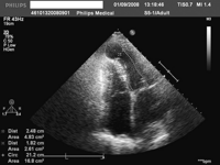
Fig. 1
Two-dimensional echocardiogram demonstrating LV aneurysm (marked round-like area in an apex of LV).
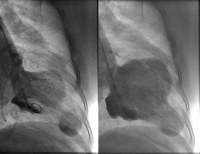
Fig. 2
LV ventriculography showed apical LV aneurysm during the ventricular systole (on the left) and diastole (on the right). The patient was successfully operated and discharged on postoperative day 9.
© European Association for Cardio-Thoracic Surgery 2008
