-
PDF
- Split View
-
Views
-
Cite
Cite
Delphine Trousse, Xavier Benoît D’Journo, Jean-Philippe Avaro, Christophe Doddoli, Roger Giudicelli, Pierre Antoine Fuentes, Pascal Alexandre Thomas, Multifocal T4 non-small cell lung cancer: a subset with improved prognosis, European Journal of Cardio-Thoracic Surgery, Volume 33, Issue 1, January 2008, Pages 99–103, https://doi.org/10.1016/j.ejcts.2007.09.032
Close - Share Icon Share
Abstract
Objective: T4-disease for non-small cell lung cancer (NSCLC) includes different conditions: mediastinal invasion, neoplastic pleural cytology, and multifocal disease in the same lobe; regarding the last category, no strict criteria allow to differentiate satellite nodules from synchronous multiple primary tumours. Methods: Retrospective study of 56 patients who underwent a complete resection from 1985 to 2006 of a NSCLC graded pT4N0 due to multifocal disease. A small nodule (<1 cm) closed to the primary tumour, in a same pulmonary segment with an identical histology was considered as a satellite nodule (pT4sn). Multiple tumours, sized more than 1 cm, with an identical histology, located in the same lobe but in different segment were considered as synchronous cancers (pT4sc). Results: There were 44 males and 12 females: 35 patients were graded T4sn and 21 patients T4sc. The median age was 62.5 years. The two groups were similar for sex, age, tobacco consumption, ASA score, NYHA, Charlson’s index, spirometric parameters, cardiovascular comorbidity and history of previous extra-thoracic malignancies. All had a complete anatomic resection with mediastinal lymphadenectomy. Thirty-day mortality rate was 3.6%. Overall 5-year and 10-year survival rates were 48.2% and 29.9%, respectively. There was a non-significant trend for a worse survival in T4sn group patients when compared to that of T4sc group patients: 42.9% vs 52.3% at 5 years, and 25% vs 34.9% at 10 years (p = 0.62). Conclusions: Multifocal T4 stage IIIB disease is a heterogeneous category where overall prognosis is far better than those of other T4 subgroups. Survival rates associated with pT4sn and pT4sc look roughly similar because of the small size of the subgroups usually submitted to comparison in most series. In the present experience, respective survival figures diverge, suggesting different biological behaviours.
1 Introduction
The current TNM staging system for primary lung cancer [1] remains imperfect especially for the T4-disease. Indeed, many different entities with distinct outcomes are included in this group (multiple primary cancer in the same lobe, satellite pulmonary nodules, mediastinal invasion or neoplastic pleural effusion). Some of them could be cured by surgery, i.e. multifocal synchronous primary cancers, while others would only require medical treatment (neoplastic pleural cytology).
The definition of such T4-multifocal disease is critical because no clear criteria to distinguish primary synchronous lung lesions from intrapulmonary satellite nodules or pulmonary metastasis in the same lobe have been established so far. Although these three different entities could possibly have different prognosis, no therapeutic guidelines exist to manage those patients specifically. As previously reported some T4-subgroups may have a surprisingly good prognosis after complete surgical resection [2], similar to that of a single primary lung cancer, and seem misclassified as stage IIIB [3].
After a cautious distinction between satellite nodules and multiple synchronous disease in a same lobe, we propose in this study to compare overall long-term survival after complete resection of these two T4-groups of patients. Aware that staging the disease is the first step of any treatment, the issue was also to clarify the current TNM Classification to make the future scheduled revision of the International Staging System for Lung Cancer more accurate for multiple NSCLC.
2 Materials and methods
From July 1985 to December 2006, 143 primary lung tumours resected in our thoracic surgical department were staged pT4 because of the presence of a multifocal disease. Eighty-one patients had intrapulmonary satellite nodules (pT4sn) around the original tumour, while 62 patients had a synchronous primary lung cancer in a same lobe (pT4sc). We have considered as satellite nodules small tumours, with an identical type cell, sized less than 1 cm, close to the primary cancer in the same pulmonary segment. On the other hand, synchronous tumours were defined as multiple tumours of different histology or, if identical, sized more than 1 cm and originating from a different pulmonary segment of the same lobe of the index tumour [2]. Our patient charts review found 6 additional patients with a tumour sized less than 1 cm and located at different segment of the same lobe, but no patient with two tumours sized more than 1 cm at the same segment of the same lobe. The 6 aforementioned patients were not included in the analysis. All patients included in the study had an anatomic lung resection associated with a mediastinal lymphadenectomy. We mainly studied the 56 patients with a pT4N0 disease, and briefly analysed separately the 67 patients with invaded lymph nodes given the attached pejorative prognostic meaning. We excluded 14 patients: those with synchronous tumours of different histology, in coherence with the group of the patients with satellite nodules, as well as those with pure bronchioloalveolar carcinoma or small cell lung cancer because of their specific behaviours.
2.1 Patients
Extracted from our prospectively filled database, 56 consecutive pN0 patients were finally reviewed in this retrospective study carried out from 1985 to 2006. Thirty-five patients were staged pT4N0 due to the presence of solitary (n = 18) or multiple (n = 17) satellite nodules and constituted the pT4sn group. In all cases, satellite pulmonary nodules were fortuitously discovered during the histopathological examination. Twenty-one patients presented with multiple synchronous primary lung cancers in the same lobe and constituted the pT4sc group. In all cases, synchronous tumours were visible before surgery on the chest CT-scan.
There were 44 males (78.6%) and 12 females (21.4%). The median age was 62.5 years and ranged from 44 to 80 years. Most of the patients were current smokers (39.3%) or ex-smokers (48.2%). The mean tobacco consumption was 37 (±28.7) packs/year (range 2–165). After clinical examination, the American Association of Anaesthetists (ASA) score and Charlson’s co-morbidity index were evaluated for all patients considering their overall fitness and co-morbidities. The mean ASA score was 1.86 (±0.9) while the mean Charlson’s index was 2.7 (±1.7), ranged from 0 to 7.
Cardiac or vascular morbidities were present in 23.2% of the patients. Previous extra-thoracic malignancies, proved to be in complete remission at the time of diagnosis of the lung cancer, were related in 14.3% of the group (n = 8). Chronic obstruction pulmonary disease (COPD) was found in 21.8% of them (n = 12) according to the GOLD guidelines [4]. Mean forced expiratory volume in 1 s (FEV1) was 72.1% of predicted (Table 1 ).
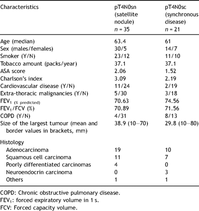
Characteristics of patients operated on for T4 multifocal NSCLC (n = 56)
2.2 Preoperative work-up
All patients due to have a resectable primary lung cancer underwent a chest X-ray, a chest CT scan, a bronchoscopy and pulmonary function tests. Some patients had further investigations, i.e. a brain CT scan (n = 49, 87.5%) and 19 (33.9%) had a bone scintigraphy.
2.3 Surgery
In operable patients, a complete anatomic resection of the tumours was performed and has been considered as the optimal therapeutic option. At least a segmentectomy, more often a lobectomy, and sometimes a pneumonectomy were done. According to oncological principles and in order to proceed with a curative approach, the surgical procedure included a mediastinal lymphadenectomy, responding to the following criteria: at least the removal of 10 lymph nodes including two or more ipsilateral mediastinal stations explored [5].
2.4 Staging
All tumours were staged after the final pathologist’s report of the specimen, using the 1997 revisions in the International System for Staging Lung Cancer [1]. Each patient was then re-staged on the basis of the index tumour disregarding the satellite nodules in the pT4sn group or smallest tumours in the pT4sc group.
2.5 Statistics
The statistical analysis was performed by using the SPSS version 13.0 software package (SPSS Inc., Chicago, IL). Quantitative variables were expressed as mean ± standard deviation (SD). Characteristics of the patients between the two groups were compared using the Pearson’s χ2 test for qualitative values and the Wilcoxon test for quantitative variables. Survival was calculated using the Kaplan–Meier method, including the operative mortality. The log-rank test was used to point out potential differences on survival among the groups. A Cox proportional hazards model was fit to examine and adjust for any explanatory variables. Forward stepwise procedure was used to select the variables with the greatest prognostic value (p < 0.05).
3 Results
3.1 Histopathology
Histology of the primary tumour was known preoperatively in 28.6% of the patients (n = 16). Cell types such as adenocarcinoma and squamous cell carcinoma were the most frequent (Table 1). The mean size of the index tumour was 34.4 mm, with 30 patients in whom the index tumour size exceeded 30 mm.
3.2 Surgery
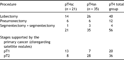
All patients (n = 56) underwent a complete R0 resection. Forty patients had a lobectomy (71.4%), including 2 who had an angioplastic and bronchoplastic sleeve lobectomy. Twelve patients had a pneumonectomy (21.4%) and 4 had a segmentectomy (7.1%) (Table 2 ). A mean of 15 (±9.3) lymph nodes were removed, exploring a mean of 3.8 (±1.7) mediastinal lymph node stations.
3.3 Staging
After re-staging, referring to the index tumour, 18 patients were found to have a pT1 disease and 37 patients with a pT2 disease, and 1 had a pT4-disease (mediastinal invasion). Among the 35 pT4sn group patients, 7 were re-staged as pT1 and 28 as pT2. Among the 21 pT4sc group patients, 11 were re-staged as pT1 disease and 9 as pT2 disease (Table 2). The distribution of T1 and T2 tumours in each group was statistically different (p < 0.01).
3.4 Treatment strategy
Three patients had induction chemotherapy. Nine patients had an adjuvant treatment: chemotherapy in 7 and radiotherapy in 2.
3.5 Outcome and long-term survival
The postoperative morbidity rate was 28.6%. Postoperative complications consisted of atelectasis (n = 8), prolonged air leak (n = 4), pneumonia (n = 3), cardiac arrhythmia (n = 2), and bronchial stump fistula (n = 1). Three patients required intensive care in the postoperative period. At the end of the study, 35 patients (62.5%) were dead.
The 30-day and 90-day mortality rates were 3.6% (95% confidence interval [CI]: 0.6–13.4) and 5.4% (95% confidence interval [CI]: 1.4–15.8). Two-year, 5-year and 10-year overall survival rates for all pT4N0 multifocal disease were 63.4% (95% [CI]: 50.7–76.1), 48.2% (95% [CI]: 34.9–61.5), 29.9% (95% [CI]: 16.2–43.6), respectively. Median survival was 45.4 months (95% CI: 8.6–82.5). The mean follow-up time after surgical resection for the whole group was 51.5 (+45.9) months. Patterns of recurrence during follow-up were as follows: 15 patients with local and 8 with distant recurrences, 33 patients dead or alive presumed free of disease.
Comparing the two groups, 5-year survival rates were 42.9% (95% [CI]: 21.7–64.1) for pT4sc and 52.3% (95% [CI]: 35.2–69.4) for pT4sn. Ten-year survival rates were 25% (95% [CI]: 5.2–44.8) for pT4sc and 34.9% (95% [CI]: 16.9–52.9) for pT4sn. The survival difference was not statistically significant (p = 0.62) (Fig. 1 ).
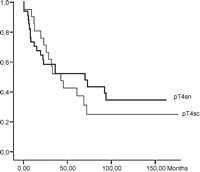
Overall survival comparing pT4N0 satellite nodules to pT4N0 synchronous cancers.
Neither sex (p = 0.22) nor age (p = 0.11) were found to impact overall survival. Comorbidities as smoking status (p = 0.67), cardiovascular disease (p = 0.56) or COPD (p = 0.82) at time of surgery did not influence survival. Postoperative morbidity was equally ranged between the two groups without significant link on outcome (p = 0.4).
Survival was not significantly affected by the histological type of both tumours (p = 0.28) nor was to the awareness of malignancy before surgery (p = 0.16). In T4sn patients, no impact on overall survival was disclosed considering the number (1 vs >1) of satellite nodules (p = 0.11).
The stage of the main tumour was the only factor detected to be a significant prognosticator both in T4sc and T4sn patients (p = 0.03) (Figs. 2 and 3 ).
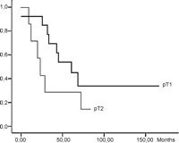
Overall survival in the pT4sc group considering the size of the biggest synchronous tumour.
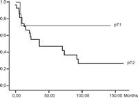
Overall survival of the pT4sn considering the size of the main tumour, disregarding the satellite nodules.
4 Analysis of pT4N+ patients
During the same period of time, 67 additional patients were operated on for a pT4snN+ (n = 35) or a pT4scN+ disease (n = 32). Mean tumour size values were 51.2 mm [20–120] and 42.7 mm [15–130], respectively. In this group of 67 patients, the median survival was 22.8 months (95% CI: 9.9–35.8), and overall 5-year and 10-year survival rates were 30.1% and 18.3%, respectively. In the pT4snN+ group, overall 5-year and 10-year survival rates were 40% and 25%, respectively. In the pT4scN+ group, 5-year and 10-year survival rates were 21.7% and 13%, respectively. This difference of survival was at the border of statistical significance (p = 0.08). When pooling N0 and N+ patients, overall 5-year and 10-year survival rates were 47.5% vs 34.9% and 29% vs 16.3% in T4sn vs T4sc patients (p = 0.39). At multivariate analysis, only the N+ nodal status influenced negatively the survival of the 123 patients with a multifocal T4-disease (HR = 0.59, p = 0.031). In contrast, the size of the index tumour (more than 30 mm vs less than or equal to 30 mm) did not impact on survival significantly (HR = 0.76, p = 0.26).
5 Discussion
Multifocal primary lung cancer is a critical issue for diagnosis, staging and treatment. Firstly, the problem remains to distinguish synchronous primary lung tumours from satellite pulmonary nodules and finally intrapulmonary metastasis. Previous published studies did not establish a clear differentiation between satellite pulmonary nodule and synchronous primary tumours with identical histology in a same lobe, because of the absence of a consensual definition [6–9]. Deslauriers et al. defined satellite nodules as ‘as well-circumscribed accessory carcinoma foci clearly separated from the main tumour but with identical histologic characteristics’ [9]. This explanation remains vague with respect to the number of lesions, the size limit, and the required margins from the original tumour. Some publications have considered that any additional tumour of identical histology in a same lobe consisted of satellite nodule while those located in different lobes were synchronous tumours [10,11]. Others studies did not care about the location of the additional tumour, considering all as satellite lesions and excluding the possible event of two independent primary cancer [7,8,12–14]. Finally, mixed groups of satellite nodules, synchronous primary cancers or intrapulmonary metastases have often been improperly compared previously, and some authors concluded on a similar fate [3,15].
We opted for a pragmatic definition of both entities, based on pathological clues. We aimed to investigate on their respective prognosis because satellite nodules are generally regarded as a local but metastatic process in nature [7–9,15], whereas synchronous multiple tumours represent a random clinical occurrence, the reality of which is not taken into account by the current TNM staging system at all [2]. Ten years ago, some other authors, interested in intrapulmonary metastasis in a same lobe also reported substantial survival rates tending to 49% 3 years after surgery, and that although additional pulmonary nodules in a same lobe were bad prognosticators, overall survival remained fairly correlated to the T-stage of the index tumour [8,11]. Similar patterns were recently reported by Port et al. [3]. Accordingly, we showed that the pT status of the index tumour impacted significantly on the outcome. In agreement with others, we support the contention that no additional preoperative work-up nor a different therapeutic approach is required for this subgroup of T4-disease [11].
In the present study, we aimed to focus on those patients selected on the basis of the absence of lymph nodes metastases in order to fit with the hypothesis of only a local disease. In N+ patients, the mean size of the index tumour was significantly higher than that of the index tumour of N0 patients, thus suggesting a correlation between the tumour size and the existence of nodal metastasis. As in other T categories, the N status influenced most survival, and was the only independent prognosticator of poor survival in this group. Like others, we observed that the surgical management for a T4-multifocal disease provided a better prognosis (42.9% 5-year survival rate for satellite nodules and 52.3% for synchronous cancer in N0 patients, 40% and 21.7%, respectively in N+ patients) than for other stage groupings within T4-disease that cover a poorer outcome, with a 5-year survival ranging roughly from 0% to 20% [1,3,11,14–16]. For this reason, multifocal disease should not be scaled as stage IIIB, which is, in most physician minds, a synonym for a non-resectable disease. It would be preferable to grade these tumours as T3 tumours. This is all the more important as the incidence of indeterminate satellite lesions is expected to increase as the consequence of continuously improved investigational technologies. The current incidence varies from 5.9% to 16% of the patients presenting with an otherwise ‘surgical’ stage [12,13,17]. Finally, benign coincidental satellite nodules, a possible matter of confusion, are commonly detected in cancer patients [17–19].
We confirmed that the occurrence of a multifocal lung cancer in a same lobe, independent of the number of satellite nodules or its cell type, has a noticeably poorer prognosis than a single NSCLC [9,11,14,15]. However, a substantial 5-year survival approximating 50% in N0 patients and 30% in N+ patients was reached, which is in line with published data [3,16]. No significant difference on survival was pointed out between pT4sn and pT4sc, both in N0 patients as well as in N+ patients, thus justifying their grouping in the same prognostic category. However, the respective survival figures in N0 patients diverged evidently after 3 years resulting in markedly different 10-year survival rates. In N+ patients, the difference of survival was close to being significant. Finally, T4sn patients tended to have a paradoxically better outcome than that of T4sc patients, despite an imbalanced distribution of T1/T2 index tumours at the disadvantage of the T4sn group. These features suggest different biological behaviours.
6 Conclusion
Multifocal T4-disease targets various and not well-defined conditions. We propose to distinguish synchronous tumours from satellite nodules on the basis of pathological clues easily accessible on the operative specimen. Even if speculative, it is highly probable that these 2 entities follow different biological phenomenon. Their grouping into the same prognostic category remains valid, as it is always the case when the issue is uncommon and statistics are based on small retrospective series only. Anyway, the attached prognosis after resection is far better than that of other T4 categories, and does not reflect at all the dismal outcome of stage IIIB disease, and justifies surgery as the pivotal part of their treatment. It would be opportune to move these conditions down in lower stages at the occasion of the coming sixth revision of the TNM classification in 2007.
References
Author notes
Presented at the 15th European Conference on General Thoracic Surgery, Leuven, Belgium, June 3–6 2007.


