-
PDF
- Split View
-
Views
-
Cite
Cite
Alfredo N.C. Santana, Mauro Canzian, Rafael Stelmach, Fabio B. Jatene, Placental transmogrification of the lung presenting as giant bullae with soft-fatty components, European Journal of Cardio-Thoracic Surgery, Volume 33, Issue 1, January 2008, Pages 124–126, https://doi.org/10.1016/j.ejcts.2007.10.007
Close - Share Icon Share
Abstract
A 44-year-old man presented with progressive dyspnea and a previous pneumothorax. Chest CT scan showed a mediastinal shift due to giant bullae containing soft tissue and fatty components in the left lower lung lobe, and a right upper lung lobe partially collapsed. The pulmonary function tests revealed forced vital capacity (FVC) 53% (of the predicted) and forced vital capacity in 1 s (FEV1) 52%. Then, resection of the lower lobe was performed with intention to prevent other pneumothoraxes and to revert the upper lobe collapse. The pathological examination showed a placental transmogrification of the lung (PTL). One month after the surgery, the patient was asymptomatic, the pulmonary function tests normalized and the upper lobe was well expanded. In conclusion, we described the first CT finding of soft tissue and fatty components within the PTL-related bullae, and the PTL should be considered in the differential diagnosis of pulmonary lesions with soft-fatty and air components.
1 Introduction
The placental transmogrification of the lung (PTL) is a very rare benign lesion first described in 1979, whose pathophysiology is still not completely understood [1,2]. In relation to the word transmogrification, it comes from transmogrify (verb), which has an unknown etymology and suggests a strange or preposterous metamorphosis, or a great change often with grotesque effect. In the literature (PubMed database), there are only 24 reported cases of PTL. The patients ranged in age from 25 to 60 years and typically presented with dyspnea, thoracic pain and/or pneumothorax. Radiographically, PTL usually presents as a bullous lesion and rarely as a cyst or nodule, and surgical resection is the treatment of choice [3,4]. Grossly, PTL resembles a placenta, and microscopically it is filled with papillary structures covered by epithelial cells, like a placental villi [1–5].
The aim of this paper is to describe a case of PTL with atypical CT findings and the improvement of pulmonary function tests after surgery. In addition, we review the literature about this pathological entity.
2 Case report
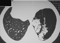
A 44-year-old man presented with a 3-year history of progressive dyspnea and one episode of left pneumothorax 6 months ago. He was a 15-pack-year smoker and had no other significant diseases. Physical examination revealed absence of breath sounds on the left hemithorax. Laboratory tests were unremarkable. Chest radiographs showed mediastinal shift to the right due to bullous lesion taking almost the entire left hemithorax. In the lower left lung lobe, a chest CT scan revealed a giant bullae with soft tissue and fatty components resembling a bunch of grapes (Fig. 1 ). In addition, the CT showed the upper left lung lobe restricted by the bullae. The pulmonary function tests (PFT) results are: FVC 1.68 l (53% of the predicted), FEV1 52%, FEV1/FVC ratio 0.82, TLC 108%, RV 233% and DLCO 51%. The room-air pulse oximetry was 92%.

Chest computed tomographic scan revealing a giant bullae with soft tissue and fatty components, resembling a bunch of grapes.
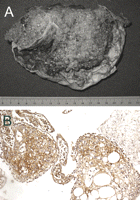
The patient underwent a left thoracotomy to resect the bullae. The idea was to prevent other pneumothoraxes and to revert the upper lobe collapse with a possible improvement in dyspnea and PFT. The bullae was resected with lower lobectomy and seemed similar to a placenta, measuring 15 cm × 14 cm × 7 cm and weighting 410 g (Fig. 2 ). The hematoxylin–eosin stain showed multiple villi composed by adipose tissue, rich vascularization, focal mononuclear infiltrate, and loose edematous connective stroma containing some cells with rounded nuclei and vacuolated cytoplasm. These cells were immunoreactive for CD-10 and vimentin. These findings characterize the PTL [1–4].

(A) Macroscopic appearance of the bullae, resembling a placenta. (B) Photomicrograph showing stromal cells strongly and consistently immunoreactive for CD-10 (×200).
One month after the surgery, the patient was totally asymptomatic. He had a room-air pulse oximetry of 97% and a new PFT showing a result totally normal. A new chest CT scan showed a centered mediastinum and the left upper lung lobe completely expanded.
3 Discussion
The original feature of our case is the first CT finding of soft tissue and fatty components within the PTL-related bullae, resembling a bunch of grapes. Radiographically, the PTL often presents as bullous lesions, as in 17 of the 24 reported cases, or still as cysts and nodules [5–7]. In addition, the mediastinal shift was present only in five previous cases [5–7]. Another important finding in our report was the significant clinical and functional improvement, such as normalization of the room-air pulse oximetry and the PFT.
Grossly, the PTL resembles a placenta or cystic lesions [1–7]. These areas are replaced or filled by gelatinous tissues described as bubbly, vesicular, grape-like or sponge-like. The differential diagnosis includes cystic adenomatoid malformation, mucinous cystic tumor of the lung, and cystic mucinous variant of bronchioloalveolar carcinoma, but the PTL can be readily distinguished from them on microscopy [2].
The hematoxylin–eosin stain shows papillary structures covered by hyperplastic pneumocytes and containing interstitial clear cells embedded in a fibrovascular core. The immunohistochemical-staining pattern is positive for CD-10 and vimentin and negative for cytokeratin, desmin, S-100 protein and smooth muscle actin [2]. As seen from the above findings, the PTL exhibits an immature mesenchymal phenotype [2].
In conclusion, although rare, the PTL should be considered in the differential diagnosis of giant bullae containing soft tissue and fatty components. Histological and immunohistochemical findings differentiate the PTL from emphysema, pleuropulmonary blastoma, sarcoma, sclerosing hemangioma and solitary fibrous tumor, suggesting that PTL probably represents a benign proliferation of peculiar interstitial clear cells with emphysema-cyst-like changes. In addition, surgical resection of the PTL may restore the physiological function of the lung.
Acknowledgements
We thank Cleidson de Araujo Rangel Junior for the English review.


