-
PDF
- Split View
-
Views
-
Cite
Cite
Rui Haddad, Multiple asymptomatic lateral thoracic meningocele, European Journal of Cardio-Thoracic Surgery, Volume 33, Issue 1, January 2008, Page 113, https://doi.org/10.1016/j.ejcts.2007.09.025
Close - Share Icon Share
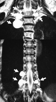
A 66-year-old woman had an abnormal chest X-ray. MRI scan showed multiple thoracic lateral meningoceles (Fig. 1 ). Meningocele is a herniation of the meninges through a vertebral column defect. Asymptomatic non-neurofibromatosis-related multiple thoracic lateral meningoceles is a very unusual finding. No treatment is necessary for this case.

Composite picture of spinal MRI scan showing 8 lateral thoracic meningoceles (white arrows), 5 in the upper and 3 in the lower paravertebral area. Meningocele should be included in differential diagnosis of posterior mediastinal cysts. There are some reports of ruptured meningoceles in the literature. This patient was not admitted to hospital, the lesion was seen in a routine chest X-ray, confirmed with CT and diagnosed by MRI and she is being followed up for 3 years.


