


Abstract
Objective: Correct staging, optimal resection type, and prognosis for non-small cell lung cancer (NSCLC) with invasion of the adjacent lobe through the fissure have seldom been reported. Methods: We retrospectively evaluated 351 completely resected NSCLC patients between 1994 and 2004. Of these, 152 patients had T2 and 139 had T3 NSCLC confined in one lobe and 60 patients had T2 NSCLC that shows a limited growth through the interlobar fissure into the adjacent lobe (NSCLC-ALI). Types of resections performed in patients who have NSCLC-ALI were: pneumonectomy in 40, bilobectomy in 10, and lobectomy plus partial adjacent lobe resection (LPR) in 10. Survival rates of all patients were determined and factors affecting the survival were evaluated by univariate and multivariate analyses. A multivariate survival analysis of NSCLC-ALI patients including the resection type as a prognostic factor was also performed. Results: Survival of the patients with NSCLC-ALI was not statistically different from those with T3 disease (p = 0.67, log rank test) but was significantly poorer than remaining patients with simple T2 disease (p = 0.049, log-rank test). T status was found as a prognostic factor at multivariate analysis too (p = 0.037). The survival of patients who underwent pneumonectomy was significantly worse than the patient group who underwent bilobectomy or LPR (p = 0.04). There was no statistically significant difference between survival of the patients who underwent LPR and the patient group who underwent pneumonectomy or bilobectomy (p = 0.16). Hospital mortality was 6.6% (4/60) and they all underwent a pneumonectomy. During follow-up there was no local recurrence encountered in patients in LPR group. Conclusions: The prognosis of NSCLC with limited invasion of an adjacent lobe was found to be similar with that of T3 tumors. A resection type lesser than a pneumonectomy may be considered in these tumors.
1 Introduction
Visceral pleural invasion appeared in the mid-1970s and remained unchanged until now as a specific entity in the TNM classification of NSCLC rendering the lesion to be T2 [1]. Tumors invading the surface of interlobar pleura are also T2. However, there is no clear definition what T factor should correspond to a tumor that invades an adjacent lobe beyond the interlobar pleura.
Resected NSCLC with invasion beyond interlobar pleura was studied to evaluate the significance of limited invasion of an adjacent lobe, to better understand its role as a prognostic factor, and to clarify the optimal resection type when encountered with it.
2 Patients and methods
From 1994 to 2004, surgical resection was performed in 337 male and 14 female patients with NSCLC in our center. Patients were evaluated retrospectively. The mean age was 55.6 ± 9.2 (32–75) years. Patients with positive surgical margin, satellite lesions, T4 tumor, superior sulcus tumor, N3 disease, or distant metastasis verified by intraoperative findings or postoperative pathologic examination were excluded from the study. Patients whose tumors were subsequently classified as small cell carcinoma or low-grade malignant tumor, as well as those who received any kind of neoadjuvant treatment, were also excluded.
Preoperative workup included routine biochemical tests, ECG, basic pulmonary function tests with or without DLCO and V/Q scan, and blood gas analysis. Moreover, all had a preoperative chest X-ray, thorax computerized tomography, fiber optic bronchoscopy, and some had thoracic magnetic resonance imaging (MRI). Cranial tomography and bone scans were also carried out in majority of cases.
Mediastinoscopy was performed in all cases except the ones with peripheral T1 squamous cell carcinomas that were histologically confirmed. Resection types included lobectomy, bilobectomy, pneumonectomy, and lobectomy plus partial adjacent lobe resection. Systematic mediastinal lymph node dissection was routinely carried out in all patients. Pathologic staging was performed according to the New International Staging System for Lung Cancer [1]. Histological typing of the specimens was done according to the World Health Organization 2004 classification [2].
T3 lesions were divided into three subgroups: bronchial (tumor in the main bronchus within 2 cm distance from the carina, but without involving it, or associated atelectasis or obstructive pneumonitis of the entire lung), mediastinal (tumors invading mediastinal pleura, pericardium, pulmonary artery, or veins extrapericardially), and peripheral (tumors invading parietal pleura or chest wall).
Patients with NSCLC invading adjacent lobe beyond interlobar pleura were studied as a separate group. Adjacent lobe invasion was defined as direct invasion of the adjacent lobar parenchyma by the tumor only a few centimeters in depth or width. Lesions showing invasion as limited only into the visceral pleura of the adjacent lobe were also included. Patients with major invasion of the adjacent lobe were excluded from this group and studied with other patients, treated with bilobectomy or pneumonectomy. Existence of invasion was confirmed with histopathologic examination in order to be included in the NSCLC-ALI group. Decision of the resection type was also based on pulmonary reserve of the patient and localization of the tumor if eligible for partial resection, in each patient. This group of patients formed the basis of this study.
The mean follow-up time was 27, 6 ± 21 (2–120) months.
2.1 Statistical analysis
Patients’ survival was expressed by the Kaplan–Meier (univariate) and Cox proportional hazards model (multivariate) analyses, using thoracotomy time as the time zero and death time, if occurred, as the end point. Differences in survival were determined by log-rank test at univariate analysis and prognostic factors having p-value less than 0.1 were included in multivariate analysis. Factors affecting the survival were determined and results were considered significant if the p-value was less than 0.05 at multivariate analysis. Hazard ratios of the prognostic factors were given with 95% confidence intervals.
3 Results
Among the patients, 152 were pathologically staged as T2, 139 as T3, and 60 as NSCLC-ALI. Mean age of the patients, gender distribution, histological types, mean tumor size, N status, and resection types of each group are listed in Table 1 .

Some characteristics and resection types of the patients
Among the NSCLC-ALI group, the primary tumor was in the right lung in 36 patients (upper lobe in 14, middle lobe in 6, and lower lobe in 16) and in the left lung in 24 patients (upper lobe in 13 and lower lobe in 11). Twenty-three (38.3%) patients had no nodal metastases (N0 disease), 29 (48.3%) had hilar–interlobar nodal metastases (N1 disease), and 8 (13.3%) had mediastinal nodal metastases (N2 disease). The extent of the pulmonary resection for the primary lesion was pneumonectomy in 40 patients, bilobectomy in 10, and lobectomy with partial adjacent lobe resection in 10. Four of the 60 patients (6.6%) died within the first 30 days in this group. They had all undergone pneumonectomy.
The cumulative 5-year survival rate for NSCLC-ALI group was 36%. Among the other patients, 5-year survival rates were 56% in T2 group and 40% in T3 group. The cumulative 5-year survival rates based on resection type was 30% in pneumonectomy and 53% in lobectomy group. Five-year survival rates were 49%, 38%, and 22% in pathologic N0, N1, and N2 groups, respectively.
The evaluated prognostic factors in univariate and multivariate analyses were age, gender, histology, tumor size, tumor differentiation, pT status, pN status, and type of resection. Of these, presence of N disease, T status, resection type, and tumor size were found to be significant prognostic indicators in univariate analysis (p = 0.0004, 0.0026, 0.0026, and 0.01, respectively, log-rank test). Among these, N disease, T status, and tumor size were also found to be significant prognostic factors in multivariate analysis (p = 0.043, 0.037, and 0.038, respectively) (Table 2 ).
Characteristics of NSCLC patients with pT2, pT3, and with adjacent lobe invasion, characteristics of their tumors and prognostic values of these variables by univariate and multivariate analysis
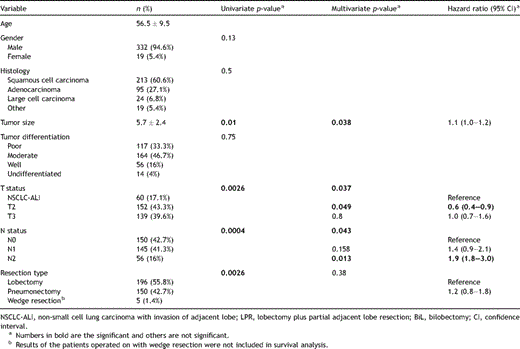
When the T status of the patients was compared, the 5-year survival rate was significantly worse in the NSCLC-ALI group than that of patients with T2 disease (p = 0.04, log-rank test), and was statistically not different from the survival of patients with T3 disease (p = 0.67, log-rank test) (Fig. 1 ). In multivariate analysis also, a statistically significant difference was found between survival rates of NSCLC-ALI and T2 groups (p = 0.049), but no significant difference was found between survival rates of NSCLC-ALI and T3 groups (p = 0.8) (Table 2).
Survival curves of NSCLC patients staged as T2, T3, and with adjacent lobe invasion (ALI, NSCLC with adjacent lobe invasion).
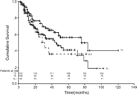
Of the patients with T3 tumors, 46 were in bronchial, 20 were in mediastinal, and 73 were in peripheral subgroups. The 5-year survival rates of patients with bronchial, mediastinal, peripheral, and interlobar pleura were 55%, 36%, 35%, and 36%, respectively. The difference between interlobar pleural invasion group and T3 subgroups was not statistically significant (p = 0.56) (Fig. 2 ).

Survival curves of NSCLC patients with interlobar pleural invasion and with T3 subgroups (ALI, NSCLC with adjacent lobe invasion).
The survival of patients who had invasion of the other lobe through the interlobar pleura was stratified according to the type of resection (pneumonectomy, bilobectomy, and LPR groups). When compared with log-rank test, survival difference between pneumonectomy or bilobectomy group versus LPR group was not statistically significant (p = 0.16, log-rank test). However, statistically significant (p = 0.04, log-rank test) survival difference between bilobectomy or LPR group versus pneumonectomy group was found (Fig. 3 ). The effect of resection types on survival of NSCLC-ALI group was evaluated with histological tumor type and N status by multivariate analysis (other prognostic factors were not included due to their limited patient numbers). Resection type was not found effective on survival at multivariate analysis. Survival of the patients in pneumonectomy group versus that of bilobectomy or LPR group was also not different (p = 0.053).
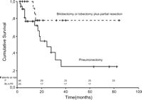
The survival curves of bilobectomy or lobectomy plus partial resection group vs pneumonectomy group (P, pneumonectomy; BL/LPR, bilobectomy or lobectomy plus partial resection).
Postoperative mortality rates were found to be 10%, 0%, and 0% in pneumonectomy, bilobectomy, and LPR groups, respectively. There was no local recurrence observed in patients operated upon with LPR during follow-up.
4 Discussion
Revisions in the international lung cancer staging [1] have not addressed the tumors invading interlobar pleura and adjacent lobe. In this study, we compared the outcome of patients with NSCLC with such an invasion and that of other NSCLC patients staged as T2 and T3, in order to delineate the aggressiveness of these tumors, and to clarify what their T status should be. Another end point of this study was meant to be the investigation of the significance of the resection in case of small invasion of an adjacent lobe beyond interlobar pleura by NSCLC.
There have been few reports regarding prognosis after resection of lung carcinomas invading the interlobar pleura and adjacent lobe. Okada et al. [3] suggested that patients with lung cancer invading interlobar pleura and other lobes have prognosis like patients staged as T3 NSCLC. On the contrary, Miura et al. [4] did not find any significant survival difference between patients with NSCLC invaded interlobar pleura and with T2 lung cancer. The latter finding was confirmed by Nonaka et al. [5] in case of squamous cell lung carcinoma; however, they shared poorer survival in patients with adenocarcinoma. Our findings were parallel with Okada’s as our NSCLC-ALI group of patients had poorer prognosis than T2 patients. Despite the limited number of patients, similarity of the prognosis of patients with NSCL-ALI to that of the T3 lesions supports the idea that these patients should be classified as having T3 tumors. Our experience is that tumors invading another lobe beyond interlobar pleura behave more aggressively than T2 tumors, like tumors invaded adjacent tissues having other lymph flow system as parietal or mediastinal pleura.
Controversy exists over the treatment of tumors with invasion through the fissure. Since extensive resection of malignant tumors with their lymphatic drainage is the accepted principle to prevent dissemination, curative resection can only be anticipated in lesions traversing the fissure if all of the involved tissue is anatomically removed. Theoretically this requires at least bilobectomy or pneumonectomy. However, as in previous studies, we did not observe statistically significant survival differences between patients who had undergone anatomical resection (pneumonectomy or bilobectomy) versus LPR [3,5]. Furthermore, we found that survival of the patients who underwent pneumonectomy was worse than others, in univariate analysis, but only approached significance in multivariate analysis (p = 0.053), probably due to number limitation. Loco-regional recurrence risk should be lower in patients who received anatomical resection than those with partial resection theoretically. But, we did not observe loco-regional recurrence and poorer prognosis in our patients treated with LPR, as reported in other reports [3,5]. Besides, the patients who underwent pneumonectomy had higher mortality rates. We therefore postulate that complete removal of the adjacent lobe would provide no survival advantage at the expense of performing a more complicated operation with higher morbidity and mortality. Additionally, in NSCLC patients who supposed small invasion of adjacent lobe intraoperatively, invasion cannot be confirmed histopathologically and unnecessary pneumonectomy may be done erroneously. Considering its higher morbidity, mortality, lack of survival advantage, in cases of NSCLC-ALI, performing a lobectomy with wedge resection of adjacent lobe or a bilobectomy on right lung may be preferable over a pneumonectomy.
In summary, it is our belief that NSCLC invading through the fissure into the adjacent lobe is better staged as T3 rather than T2 and it is our experience that pneumonectomy for resection of such tumors is not justifiable unless there are other technical reasons that necessitate such extensive resection. We would still like to address the limitation of this paper due to small number of subjects and encourage further prospective reports on the subject in larger patient populations.
References
Author notes
Presented at the 15th European Conference on General Thoracic Surgery, Leuven, Belgium, June 3–6, 2007.
