-
PDF
- Split View
-
Views
-
Cite
Cite
Dominique Gossot, Ricard Ramos Izquierdo, Philippe Girard, Jean-Baptiste Stern, Pierre Magdeleinat, Thoracoscopic resection of bulky intrathoracic benign lesions, European Journal of Cardio-Thoracic Surgery, Volume 32, Issue 6, December 2007, Pages 848–851, https://doi.org/10.1016/j.ejcts.2007.09.003
Close - Share Icon Share
Abstract
Background: Video-assisted thoracic surgery (VATS) is used for the diagnosis and treatment of some mediastinal lesions. However, large-size tumours are usually approached by thoracotomy or sternotomy. We report our experience of a full thoracoscopic approach for bulky intrathoracic lesions. Methods: From November 2002 to March 2007, 14 patients with a bulky intrathoracic mass were referred for resection. The study group consisted of eight females and six males with a mean age of 44 years (range: 13–74). We defined as bulky a mass with a minimal cross-sectional diameter equal to or larger than 50 mm, as measured on the specimen by the pathologist. Results: Thoracoscopic resection was completed in all patients. In 4 cases, the mass originated from the pleura, and in 10 cases from the mediastinum. The larger diameter of the lesion ranged from 50 mm to 160 mm, with a median of 90.2 mm. Operative time, calculated from insertion of the first trocar to skin closure, ranged from 40 to 190 min (mean: 102). Mean chest drain duration was 2.1 days (range: 1–4 days) and the mean hospital stay was 4.3 days (range: 3–11 days). There were no major postoperative complications. The final pathological diagnoses were the following: solitary fibrous tumours of the pleura (4), benign thymic cysts (2), teratomas (2), bronchogenic cyst (1), benign thymoma (1), pleuropericardial cyst (1) and benign neurogenic tumours (3). Conclusions: With experience and use of appropriate instrumentation, resection of bulky intrathoracic lesions by thoracoscopy is feasible and safe. It should be considered as a reliable alternate for tumours that are benign and most often asymptomatic.
1 Introduction
Video-assisted thoracic surgery (VATS) is widely used for the diagnosis of mediastinal masses [1,2]. It is also considered as the technique of choice for removing small benign mediastinal lesions, such as neurogenic tumours [3], bronchogenic cysts [3] and some pleural masses [4]. However, when preoperative computed tomography (CT) or magnetic resonance imaging (MRI) reveals a large intrathoracic lesion, the surgeon feels more comfortable and more confident with a conventional open approach. In selected patients, a thoracoscopic approach is feasible and might be preferable. We have reviewed our series of bulky intrathoracic tumours which were removed by VATS.
2 Patients and methods
We defined as bulky a mass with one of the cross-sectional diameter being equal to or larger than 50 mm. The measures were taken on the specimen before fixation and were sometimes smaller than those marked on preoperative CT, especially for cystic lesions whose liquid content was evacuated during the procedure. From November 2002 to March 2007, 14 patients with a bulky intrathoracic mass were operated on by VATS. There were eight females and six males with a mean age of 44 years (range: 13–74). Preoperative symptoms were observed in four patients: atypical thoracic pain in 3 and dyspnea in 1. All patients underwent chest roentgenography and CT of the chest. Five patients had MRI and two patients underwent CT-guided fine-needle aspiration biopsy. The study of serum tumour markers (alpha-fetoprotein and beta HCG) was negative in all patients with anterior mediastinal mass.
All patients were operated on under general anaesthesia with double-lumen endotracheal intubation. All were positioned in lateral decubitus, except the patient presenting with a suspicion of benign thymoma who underwent a radial thymectomy via a bilateral thoracoscopy in supine position. Three to five ports were used: one for a 10 mm–0° thoracoscope and the working ports for endoscopic instruments, ultrasonic dissector and/or endostapler. Adequate positioning of the endoscope was chosen after thorough examination of the CT, so that the tip of the endoscope is remote from the tumour and to have optimal space to manoeuvre instruments. When the tumour was of cystic content, it was partly evacuated. A purse-string suture was placed and the cyst was then punctured using a sharp tip trocar connected to suction. The trocar was then removed while the purse-string was tightened. If necessary, i.e. when the lung hampered dissection despite deflation, optimal exposure was achieved by retracting the lung with an atraumatic 3 mm grasping forceps. These allow efficient lung retraction with minimal parietal trauma.
The main concern during dissection was keeping an accurate view on the phrenic and/or vagus nerve and on all aspects of the tumour because its large-size sometime prevented a look around or behind it. It was sometimes necessary to switch from a 0° to a 30° viewing telescope.
All specimens were placed into an endobag and one of the ports was enlarged for retrieval. Usually we chose to make the larger incision into the axilla, both because of cosmetic reason and because the ribs spread apart with ease at that level, thus facilitating extraction. None of the specimen was morcellated. Only the muscular layer was closed and no intercostal suture was placed.
Duration of the procedure was defined as the time elapsed from trocar insertion to skin closure.
3 Results
The preoperative CT showed the tumour as a solid lesion in seven cases and a cystic lesion in 7. The lesion was right-sided in seven patients and left-sided in seven patients.
In 4 cases, the mass originated from the pleural, and in 10 cases from the mediastinum, either posterior in 4 cases (3 neurogenic tumours and 1 bronchogenic cyst), or anterior in 6. The larger diameter of the lesion ranged from 50 mm to 130 mm, with a median of 81.5 mm.
Thoracoscopic resection was completed in all patients. Neither mortality nor postoperative complications were recorded, except a minor parietal haematoma at the extraction site. Operative time ranged from 40 to 190 min (mean: 102 min). Mean chest drain duration was 2.1 days (range: 1–4 days) and the mean hospital stay was 4.3 days (range: 3–11 days). All but three patients had a postoperative stay inferior to 4 days. One patient stayed 7 days because of associated laminectomy for a dumbbell tumour and one old patient suffering from chronic bronchitis stayed 11 days because of postoperative hypoxia without pulmonary of pleural complication. These two patients with prolonged stay explain that the mean hospital stay is 2 days longer than the mean drainage duration. All patients were reviewed as outpatients at 1 month after discharge from the hospital and the control chest X-ray was always normal.
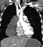
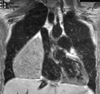
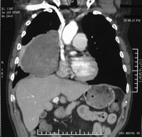
The final pathological diagnoses were the following: benign thymic cysts (2), teratomas (2) (Figs. 1 and 2 ), bronchogenic cyst (1), benign thymoma (1), pleuropericardial cyst (1) and benign neurogenic tumours (3), and solitary fibrous tumours of the pleura (4) (Fig. 3 ).

Example of giant benign teratoma in a 13-year-old girl: preoperative computed tomography.

Giant benign teratoma in a 26-year-old female patient: preoperative MRI.

Large benign solitary fibrous tumour of the pleura: preoperative computed tomography.
4 Discussion
Although many publications mentioned thoracoscopy as the standard approach for many different types of intrathoracic tumours, the issue of critical size has so far not been discussed. To our knowledge, the largest intrathoracic tumour that was removed thoracoscopically was a giant lipoma whose diameters reached 18 cm × 15 cm × 5 cm [5]. The authors combined VATS and a sternum lifting method to gain space. Few of the reported series give information about the diameter of the removed lesions and most surgeons consider that giant tumours should preferably be removed through an open approach. However, several factors should be considered: (1) most patients are young, healthy and symptom free. The intrathoracic lesion is usually discovered in a routine screening and indication for surgery may seem questionable because the long-term evolution is not clear. It is therefore important to minimise chest trauma and its inherent consequences. The extraction incision that is done at the end of the procedure cannot be compared to a thoracotomy in terms of cosmetic result, pain and sequelae since no rib spreader is used and no intercostal suture is necessary [6]. (2) By using technical tricks, appropriate instrumentation and outstanding video-imaging equipment, many obstacles can be overcome.
In a series of bronchogenic cysts, Martinod et al. stressed that their mean size was smaller in the group of patients whose cyst was removed by thoracoscopy (4.3 cm) than in the group of patients who had conversion to thoracotomy (6.1 cm) [7]. For cystic tumours, we made the choice of puncturing the cyst and aspirating its fluid content, thus facilitating mobilisation and dissection. We have used this technique for some cystic teratoma, but in the latter it can only partly deflate the cyst which contains solid parts. A similar technique has been described by Iwasaki et al., using a cannula and a balloon cholangiography catheter connected to a syringe [8] while other Japanese surgeons used another type of balloon [9]. According to some authors, VATS should be the gold standard for the removal of solitary fibrous tumours of the pleura even though an additional incision is necessary for specimen extraction [4]. A case of giant fibroma that was removed by VATS has been reported [10]. There are two types of pleural fibroma: those developed on the parietal pleural are usually sessile and sometimes adherent to the chest wall. These may be malignant and should rather be removed by thoracotomy when their size is significant. Conversely, fibromas originating from the visceral pleura are usually pedunculated on a unique large vascularised attachment that can be easily divided by a single firing of endostapler. Most are benign. It would therefore be useless to perform a thoracotomy for such a straightforward and rapid operation [11]. In our series, three out of the four fibrous pleural tumours were removed within less than 1 h. Even though an incision is added to extract the bulky specimen, no rib spreader is necessary and its consequences are minimal. With respect to large fibromas developed on the parietal pleura, thoracoscopic mobilisation can be difficult and may compromise a complete resection with free margins. In these cases, an open approach seems preferable.
Pericardial cysts are never removed in our practice, since they are always benign and asymptomatic. However, when they are very large, as in the case of this series, a thoracoscopic removal is reasonable and simple to perform [12].
Appropriate instrumentation is one of the keys for these operations which may be challenging. Since dissection is usually done in close contact with the phrenic and/or sympathetic trunk and/or vagus nerve, it is important not to use monopolar electrocautery [13]. Along with others [14], we favour ultrasonic shears because the risk of thermal injury is less. However, even ultrasonic dissectors should be used with care, since thermal damage or cavitation-related injuries remain possible in case of misuse [15]. Optimal visualisation of the whole operative field is greatly facilitated by high definition imaging and by the use of a deflectable thoracoscope.
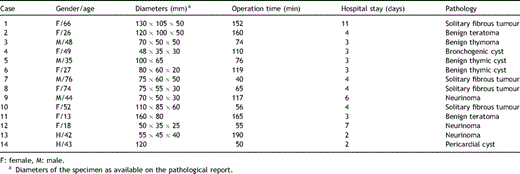
In conclusion, despite their impressive appearance, many benign intrathoracic masses can be removed without thoracotomy, provided appropriate instrumentation is used and dissection is carried out with patience and accuracy (Table 1 ).

Characteristics of patients operated on from a bulky intrathoracic tumour by video-assisted thoracic surgery
References
- teratoma
- postoperative complications
- bronchogenic cysts
- chest tubes
- cysts
- thoracic surgery, video-assisted
- thoracoscopy
- thoracotomy
- thymoma
- diagnosis
- mediastinum
- neoplasms
- pleura
- wound closure
- trocar
- thymic cysts
- solitary fibrous tumor
- intrathoracic x-ray or scan abnormality
- diameter
- medical pathologists


