-
PDF
- Split View
-
Views
-
Cite
Cite
Alexander Lembcke, Hans-Peter Dübel, Thomas Elgeti, Wolfgang Rutsch, Multislice spiral computed tomography of a malignant single coronary artery, European Journal of Cardio-Thoracic Surgery, Volume 32, Issue 5, November 2007, Page 801, https://doi.org/10.1016/j.ejcts.2007.07.015
Close - Share Icon Share
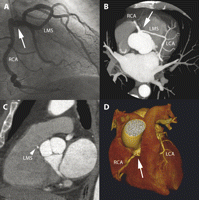
A 53-year-old man with chest pain underwent conventional angiography that demonstrated a single coronary artery originating from the right sinus of Valsalva (Fig. 1A ). However, only an ECG-gated 64-slice computed tomography definitely revealed the interarterial course of the left main segment between aorta and pulmonary trunk (Fig. 1B–D).

(A) Conventional angiography (RAO projection) of the single coronary artery with both the right and left coronary artery (RCA, LCA) originating from a single ostium (arrow). The left main segment (LMS) is a long transverse branch that crosses the base of the heart to the left side. However, the definite anatomic relationship between the left main segment and the great thoracic arteries remained unclear. (B–D) Contrast-enhanced ECG-gated 64-slice computed tomography (Panel B: maximum intensity projection; Panel C: multiplanar image reformation; Panel D: three-dimensional image reconstruction) demonstrating abnormal anatomic pattern of the coronary artery tree with a single ostium (arrows)). It became clearly evident that the left main segment courses between aortic root and pulmonary trunk (Panel C—arrowhead). This is an extremely rare type of coronary artery anomaly that has been described as potentially malignant, since a fall in coronary blood flow may occur due to squeezing or kinking of the left main segment.


