


A 67-year-old woman was diagnosed myocardial infarction due to the posterolateral artery occusion (Fig. 1A ). The chest enhanced multislice computed tomography (MSCT) scan revealed dissection of the lateral wall of the left ventricule with a 2-cm pericardial effusion (Fig. 1B). The MSCT depicted important information concerning the infarcted area (Fig. 2 ).
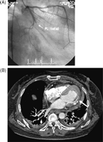
(A) Coronary angiography showing that the posterolateral (PL) artery of the circumflex coronary artery is totally occluded. (B) The chest enhanced multislice computed tomography (MSCT) shows that the lateral wall of the left ventricule is dissected (arrow).
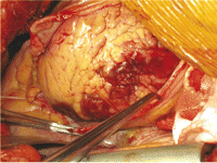
Operative findings showing necrotic myocardial tissue in the left ventricular lateral wall that was easy to expose through a lateral thoracotomy.
