


Abstract
Intrapulmonary lymph node metastasis occuring after nephrectomy for renal cell carcinoma is a particular recurrence modality. Authors report two male patients presenting with this recurrence 2 and 4 years after treatment of the primary and who underwent surgery. Surgery confirmed the diagnosis and demonstrated station 7 minimal metastases and positive pleural lavage cytology in both patients. The first patient survived 2 years and the second was alive disease free at 33 months follow-up. Such metastases probably originate from the thoracic duct. Resection confirms the diagnosis and may be part of the treatment.
1 Introduction
Mediastinal and pulmonary hilar lymph node (LN) metastases (LNM) associated with lung metastases during the course of renal cell carcinoma (RCC) are mentionned since 1965 [1]. Hilar LNM leading to RCC diagnosis was reported in 1974 [2] and those following nephrectomy for RCC in 1976 [3]. Authors’ purpose was to illustrate and comment on this particular RCC recurrence modality.
2 Case 1
A 44-year-old man underwent a left nephrectomy for RCC (grade 2, 8 × 6 × 5 cm in diameter, invading the renal capsula but without exceeding it, pT2N0M0). Two years later, a CT scan demonstrated two right hilar and one right paratracheal LN. The latter, removed by mediastinoscopy, proved to be a RCC LNM. Because the hilar LN increased (Fig. 1 ), the patient was referred to authors’ department. A right thoracotomy was performed, right hilar LNs were removed and proved metastatic (Fig. 2 ). There was no parenchymal nodule. Pleural lavage cytology was positive for RCC. Mediastinal lymphadenectomy disclosed LN micrometastasis (station 7). Remaining 4R and 2R LN were disease free. No other treatment was performed. Two right upper lobe nodules were discovered 1 year later. They were resected as well as five small nodules discovered at surgery in the middle lobe. All were metastases. Immunotherapy was performed. The patient died 9 months later from the disease.
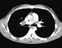
Axial chest CT view showing right hilar adenomegaly in relation with renal cell carcinoma metastasis.
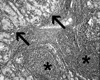
Hilar lymph node renal cell carcinoma metastasis (arrows). Lymph node is recognisable by follicular lymphoid tissue (*). (Hematoxylin eosin, ×200).
3 Case 2
A 55-year-old man underwent a right nephrectomy for RCC (grade 3, 8 × 8 × 4 cm in diameter, not invading the renal capsula, pT2N0M0). Four years later, CT scan demonstrated a right lung nodule and ipsilateral hilar LN. He was referred to authors’ department. The thoracotomy revealed a right middle lobe hamartoma. Hilar LNs removal necessitated a right upper lobectomy and confirmed RCC bulky LNM. The mediastinal lymphadenectomy disclosed two small LNMs (station 7). Pleural lavage cytology was positive for RCC. Postoperative course was uneventful. No other treatment was performed. The patient is still alive and faring well 33 months later.
4 Comment
Mediastinal and pulmonary hilar LNMs occuring during the course of RCC are not uncommon nor purely coincidental. The first observed RCC hilar LNMs were either part of the RCC clinical presentation or leading to its discovery. RCC hilar LNM presenting as recurrence following nephrectomy for RCC was first reported in 1976 [3]. Thereafter, such recurrence was regularly described and encountered after variable disease-free intervals. After Reinke et al. [3] observed first case 3 years following a nephrectomy, Wright [4] reported two cases occurring 1 and 3 years later, Merine and Fishman [5] three cases 9 months, 2 and 4 years later and others reported still longer intervals: 7 years [6], 19 years [7] and 20 years [8]. Two of these late presentations [6,7], were treated by surgery as was also treated a case of Wright [4]. To author’s knowledge, these are the only three cases comparing with authors’ and sharing the same curative therapeutic purpose.
Several hypotheses have been provided by the previous reporting authors to explain hilar and mediastinal LNMs. Wright [4] stressed the right predominance of hilar LNMs and suggested that involvement occurred by reflux from the paraaortic LNs via lymphatics of the right pulmonary ligament, a known pathway explaining the spread of lung cancer to the abdominal LNs. However, in the cases authors are reporting, intra-abdominal LNs were disease free. Hematogenous metastasis from RCC to the thorax via the inferior vena cava being considered as predominant, Seaman [9] postulated that tumour emboli to the pulmonary artery may enter the adjacent peribronchial lymphatics which then drain anterograde to the hilar nodes. This mechanism, difficult to imagine, should probably be more readily suitable in case of associated lung metastases.
Reinke et al. [3] suggested that reflux of tumour emboli may occur from the thoracic duct (TD) into the paratracheal lymphatics, bronchopulmonary lymphatics and interlobar lymphatics, as a result of lymphatic valves insufficiency.
Authors suggest that this hypothesis [3] formulated 30 years ago, is more relevant because it is supported by anatomical data. Recent works [10] on renal lymphatic drainage demonstrate that renal lymphatics constantly reach the TD at its origin, sometimes directly without crossing through intraabdominal LN. This appears to be as an important pathway for metastasing cells as hematogenous spread, the tumour cells further pouring into the blood circulation via the TD arch. Reflux from the TD may occur within the paratracheal LNs but also directly within the pulmonary hilum as reported by this anatomical study demonstrating lymphatics issuing from the right hilum and connecting with the TD at the level of station 7. Mediastinal lymphadenectomy discovering station 7 involvement in both authors’ patients may reinforce this hypothesis. Since insufficiency of the lymphatic valves should be required for reflux from the TD to occur, this hypothesis also explains the rarity of this particular manifestation. Authors have no explanation regarding the pleural lavage cytology positivity for RCC in both patients, this discovery still needing further research.
Intra-pulmonary, hilar and mediastinal LNMs without hematogenous spread are potentially resectable. The patient reported by Wright [4] survived 3 years. One of the authors’ patient survived 22 months and the second is still alive at 33 months follow-up. These results support surgery of isolated enlarging hilar LN occuring after nephrectomy for RCC. Resection must include mediastinal LN dissection. Surgery may lead to a better understanding of the disease, provide a local control and be part of a multimodality therapeutic approach including emerging targeted adjuvant therapy.
