-
PDF
- Split View
-
Views
-
Cite
Cite
Hyun Joo Lee, Young Tae Kim, Chang Hyun Kang, Joo Hyun Kim, An accessory spleen misrecognized as an intrathoracic mass, European Journal of Cardio-Thoracic Surgery, Volume 28, Issue 4, October 2005, Page 640, https://doi.org/10.1016/j.ejcts.2005.06.042
Close - Share Icon Share
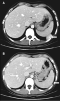
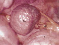
A 31-year-old woman was referred for an incidentally detected intrathoracic mass (Fig. 1). At the video-assisted surgery, we found an accessory spleen attached to the diaphragmatic surface by mesenteric pedicle (Fig. 2). It is the first case of an intrathoracic accessory spleen fed by abdominal mesenchymal vessels without trauma history and hematologic disease.

Computed tomography scan of thorax showing a homogenously enhanced mass with central feeding vessel bordering the diaphragm and the parietal pleura (A). Note a cord-like enhanced structure underneath the diaphragm, which was the vascular pedicle (B).

A round bluish-red colored mass and fatty mesenteric pedicle (thoracoscopic view). AS, accessory spleen; VP, vascular pedicle; D, diaphragm; H, heart.


