


Abstract
Objective: The ‘frozen’ elephant trunk technique allows for single-stage repair of combined aortic arch and descending aortic aneurysms using a ‘hybridprosthesis’ with a stented and a non-stented end. This report summarizes the operative- and follow-up data (mean follow-up 14 months) with this new treatment. Methods: Between 09/01 and 4/04, 22 patients (62±9 years; 9 female) with different aortic pathologies (15 aortic dissections, 7 aneurysms) were operated on after approval from the local institutional review board. The stented end of the hybridprosthesis was deployed in the descending aorta through the opened aortic arch during hypothermic circulatory arrest and selective antegrade cerebral perfusion. Results: All patients survived the procedure but one patient died of acute hemorrhage due to rupture of the false lumen in the descending aorta on the second postoperative day. Two patients required reexploration of the chest for bleeding complications. In 2 of 4 patients who developed neurological dysfunction, symptoms resolved completely. In one of them, the descending aorta was perforated intraoperatively due to misplacement of the stented end of the hybridprosthesis. In all follow-up CT-scans thrombus formation in the descending aortic aneurysm excluded by the stented end of the hybridprosthesis has been observed. Conclusions: This procedure is performed through median sternotomy and combines the concepts of the elephant trunk operation and endovascular stenting of descending aortic aneurysms. Favourable intraoperative and postoperative results during follow-up with regard to thrombus formation around the stented descending aortic segment encourage us to evaluate all patients with thoracic aneurysms extending to proximal and distal of the left subclavian artery for this treatment.
1 Introduction
Anatomy determines that the distal aortic arch and the proximal descending aorta are less accessible via median sternotomy than the upstream aortic segments. Therefore, treatment of combined pathologies of the aortic arch and the descending aorta remains a surgical challenge. In 1983, Borst described the elephant trunk operation, which facilitated staged surgery on aneurysms involving the aortic arch and the neighboring distal segments [1]. Nevertheless, staged repair carries the cumulative risks of two major surgical procedures, one through median sternotomy and the other through lateral thoracotomy [2]. This perception drove further developments in this field aimed at achieving complete repair of such extensive lesions by one single procedure [3–7]. The introduction of a new type of hybridprosthesis combining the features of a conventional vascular prosthesis and a stentgraft enables single-stage repair through median sternotomy [8]. This report summarizes our operative experience with this treatment modality including the short-term follow-up.
2 Material and methods
2.1 Patient population
Between 9/2001 and 4/2004, 22 patients were operated on after approval was obtained from the institutional review board of Hannover Medical School. Informed consent was obtained in each case. Mean patient age was 62 years (range 47–77 years). There were seven patients who were older than 70 years. Nine patients were of female gender. The majority of patients (n=11) presented with type A aortic dissection, 10 in its chronic and one with its acute variant. The second most frequent pathology (n=7) was an aneurysm proximal and distal of the left subclavian artery. A smaller cohort (n=4) presented with chronic aortic dissection type B. Additional cardiac pathology comprised severe coronary artery disease in 9 and aortic valve disease in 3 patients.
2.2 Design of the hybridprosthesis
The hybridprosthesis (‘Chavan–Haverich’ (CH) endograft, Curative GmbH, Dresden, Germany) was made of a woven vascular prosthesis with stainless steel stents affixed to the inner aspects at its distal end. The diameters of the stents ranged between 30 and 46 mm and the stents had a length of 22 mm each. The proximal portion of the hybridprosthesis was non-stented and consisted of a Dacron sleeve ready for conventional surgical handling.
The delivery system comprised a flexible 39 French (F) outer sheath, a 34 F inner sheath as well as a central pusher. Withdrawal of the outer sheath while holding the inner sheath and the pusher steady released the stented portion of the hybridprosthesis. The proximal Dacron tube was then released by pulling back both the sheaths simultaneously while holding the pusher steady. A spiral CT angiography (CTA) of the thoracic aorta was performed preoperatively to assess the extent of the aneurysm and/or dissection as well as to determine the appropriate stent graft size.
2.3 Surgery
All patients were operated using cardiopulmonary bypass. Central cannulation of the ascending aorta and the right atrium was preferred. In 3 patients who underwent resternotomy the groin vessels were cannulated before, because an ascending aortic aneurysm encroached upon the sternum. Then, core cooling was accomplished to 25 °C rectal temperature. After induction of cardioplegic cardiac arrest cardiopulmonary bypass was discontinued. The aortic arch was then opened longitudinally. Selective antegrade cerebral perfusion with cold blood at a temperature of 15° and a volume of 250–450 ml/min was initiated after ostial cannulation of the left common carotid artery and the brachiocephalic trunk. Then, the stented end of the hybridprosthesis was deployed in the descending aorta (Fig. 1 ). In the first 5 patients it was implanted over an antegradely placed super-stiff guidewire. Prompted by perforation of the aortic wall with the introducer system in a patient with a tortuous descending aorta, a through and through transfemoral guidewire technique was employed in all subsequent patients.
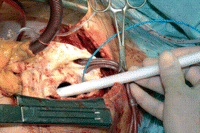
Introduction of the delivery system carrying the hybridprosthesis into the descending aorta of a patient with a combined aortic arch and descending aortic aneurysm. The aortic arch is opened during hypothermic circulatory arrest. Catheters for selective antegrade cerebral perfusion are placed in the brachiocephalic trunk and the left common carotid artery. A Fogarty catheter blocks the origin of the left subclavian artery to prevent back bleeding.
The distal landing site of the graft was at or above the 10th thoracic vertebra in all patients. After deployment, the stented portion of the prosthesis was modulated onto the aortic wall with the help of an appropriately sized balloon catheter (Medtronic, USA). Then, the non-stented Dacron graft segment was sutured circumferentially to the aorta distal to the origin of the left subclavian artery before the supraaortic branches were reimplanted enbloc into an appropriately sized window of the graft. A proximal graft-to-aortic anastomosis at any desired level of the ascending aorta completed the repair (Fig. 2 a–c). In six patients with previously implanted valved conduits a graft-to-graft anastomosis was sutured accordingly.
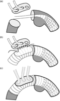
(a) The stented end of the hybridprosthesis is deployed in the descending aorta distal of the dilated segment. Selective antegrade cerebral perfusion is accomplished during hypothermic circulatory arrest. (b) After suturing the hybridprosthesis circumferentially into the descending aorta directly distal the origin of the left subclavian artery, the supraaortic branches are reimplanted into the graft as a single tissue patch. (c) The reconstruction may then be completed at any desired level of the ascending aorta while cardiopulmonary bypass is reestablished.
In 20 patients the entire aortic arch and the proximal segment of the descending aorta were replaced using this procedure. In 10 of them the ascending aorta had to be replaced additionally.
In two other patients with aneurysms limited to the proximal descending aorta, implantation of the hybridprosthesis into the descending aorta was enabled by a limited, 3–4 cm long longitudinal T-shaped incision of the aortic arch. The non-stented endograft segment was sutured circumferentially distal to the level of the left subclavian artery before the aortotomy was closed. One of the two patients required additional complete myocardial revascularization and the other aortic valve replacement for aortic valve stenosis using a biological aortic valve prosthesis.
Apart from these two patients there were 10 other patients, who required additional procedures (myocardial revascularization 8 patients, aortic valve replacement 2 patients).
3 Results
3.1 Surgery
There were no intraoperative deaths. The implantation of the prosthesis was successful in all but one patient. This patient presented with marked kinking of the descending aorta distal of the aneurysmatic segment to be excluded. Implantation was attempted over an antegradely placed guidewire. The tip of the introducer system could not be advanced beyond the kinked segment; thus perforation of the aortic wall occurred at this point, which required surgical repair and additional transfemoral stentgraft implantation to bridge the perforated and aneurysmatic segments. In all subsequent patients the transfemoral through and through guidewire technique for the implantation of the stented segment of the hybridprosthesis in the descending aorta (s.a.) was used.
In one patient with chronic aortic dissection type A the aortic wall of the false lumen directly distal to the origin of the left subclavian artery was accidentally injured during surgical preparation. Even though this laceration was sutured and the further course of the operation was uneventful, the patient died from fatal bleeding into the left hemithorax 2 days postoperatively. Autopsy revealed that exsanguination was caused by a reopening of the repaired segment.
The mean duration (±SD) of total cardiopulmonary bypass time, aortic cross-clamp time, hypothermic circulatory arrest time, antegrade selective cerebral perfusion time and time required for the deployment of the stented end of the hybridprosthesis were 239±76, 136±43, 74±19, 62±14 and 12±5 min, respectively. Reexploration for bleeding was necessary in two patients.
Four patients awoke with central neurological dysfunction. In two of them, it was transient and resolved completely before discharge. Two of the four patients had a history of cerebrovascular events with anatomical correlates in the preoperative CT scan of the brain. Documented (by direct laryngoscopy) left recurrent nerve paralysis occurred in two patients.
3.2 Follow-up
There is no late mortality after a mean follow-up of 14 months. Postoperative CT scans revealed completed thrombus formation in the perigraft space around the stented segment of the hybridprosthesis in the descending aorta in all seven patients with atherosclerotic aneurysms. The same holds true for the arch aneurysms in two patients with associated type B aortic dissections.
In the remaining 12 patients with aortic dissections in follow-up, initiation of thrombus formation in the false lumen in the descending aorta up to the level of the stents was noted in all but one patient, who exhibited a small endoleak into the false lumen at the origin of the left subclavian artery; otherwise, the false lumen was thrombosed. As the patient refused a reintervention, the exact etiology of the endoleak remains unclear.
In one patient with chronic aortic dissection type A, the stented segment of the graft could not be anchored successfully in a previously implanted thoracoabdominal aortic vascular graft. The stents slipped proximally during and after release giving rise to a distal endoleak. This was treated 2 weeks later by placing a commercially available endograft (Talent endograft, Medtronic Inc.) transfemorally, thus extending the hybrid endograft distally into the thoracoabdominal graft. At discharge, he still presented with a type III endoleak, which was found to be reduced to a tiny contrast extravasation at 6 months follow-up. As the actual aneurysm has thrombosed and its size remains constant, the patient is treated conservatively at present.
4 Discussion
Treatment of aortic arch aneuryms extending beyond the origin of the left subclavian artery either requires a two-stage approach employing the elephant trunk operation as described by Borst or an extensive single-stage procedure performed through a clamshell incision [1,3–7].
As delineated in a report by Estrera and colleagues, the staged operation carries the cumulative risks of two major procedures and the additional risk of fatal rupture during the waiting time between the two interventions [2]. Risk reduction for these patients by introduction of new alternative procedures is therefore desirable.
This report describes our experience with an approach that allows for definite treatment of lesions of the aortic arch and beyond during a single-stage procedure using a hybridprosthesis, which combines the features of a stent graft and a conventional vascular prosthesis. The stented distal segment of the hybridprosthesis is implanted into the descending aorta through the opened aortic arch under fluoroscopic control, while the proximal non-stented segment is used for conventional replacement of the upstream aorta. The procedure is performed through median sternotomy thereby facilitating additional surgery on the heart and/or the ascending aorta.
In this series of 22 patients, implantation of the hybridprosthesis was successful in all but one patient with pronounced tortuosity of the descending aorta. With the through and through transfemoral guidewire technique (s.a.) we have ever since used no malpositioning of the stented segment of the hybridprosthesis occurred.
The conventional elephant trunk operation necessitates a reoperation through lateral thoracotomy, because the perigraft space around the elephant trunk remains perfused thereby promoting further aneurysmatic dilatation of that aortic segment [2,6,7]. As opposed to this, the ‘frozen’ elephant trunk technique as described here allows for progressive thrombus formation in the perigraft space in the descending aorta up to the level of the stents. Widening of the descending aorta has not been observed in our patients, regardless of the thrombus formation being complete or incomplete. This observation suggests, that the stents within the distal segment of the hybridprosthesis are effective in preventing retrograde flow into the aneurysm.
Due to the necessity of mounting the hybridprosthesis into an adequately flexible and thin delivery system, we utilized Dacron (0.36 mm in width) which is thinner than the Dacron used in conventional aortic surgery. In addition to pretreatment with collagen during fabrication of the hybridprosthesis, extra sealing of the graft with fibrin glue was necessary in some cases to render the graft completely hemostatic.
While the periopertive mortality rate of 4.5% is well acceptable, the incidence of stroke in 4 out of 22 patients remains a concern, even though in two of the four patients symptoms resolved completely and two had had previous cerebrovascular incidents. The circulatory arrest time of 70 min in average has to be regarded as a risk factor for stroke despite the routine use of selective antegrade cerebral perfusion [9]. Deployment of the stented segment of the hybridprothesis currently takes 15–20 min. Further reduction of this time is therefore desirable, because this will shorten the phase of circulatory arrest thereby lowering the central neurological risk [10].
No patient in our series developed paraplegia as a result of spinal cord injury. Even though this result is in line with those of endovascular stent grafting of descending aortic aneurysms it is still intriguing in view of the significant paraplegia rates reported with open surgical approaches similar to ours [11–14]. One aspect that could have reduced the risk of spinal cord injury in our series was keeping exclusion criteria with regard to the extent of descending aortic aneurysm to be treated together with a stent graft fabrication, that was adjusted specifically to the patients’ individual pathology. Therefore, this treatment was limited to patients with aneuryms involving only the first half of the descending aorta regardless of their size. The proximal extent of the aneurysm with regard to arch involvement was not critical, because the quality of the anastomosis distal to the origin of the subclavian artery is rather depending on the condition of the aortic tissue than on the actual aortic diameter at that segment.
In the two patients with aneurysms limited to the descending aorta, other options such as retrograde or isolated antegrade stent graft implantation with or without transposition of the subclavian artery would have been conceivable treatment alternatives to our approach. We believe, however, that the favourable results that can be obtained with circulatory arrest at moderate hypothermia and selective cerebral perfusion in surgery involving the aortic arch well justifies the technique described here [9,10]. The use of the hybridprosthesis enables safe anchoring of its proximal vascular graft segment by a circumferential, hand-sewn anastomosis distal to the origin of the left subclavian artery on the expense of a probably somewhat extended circulatory arrest time, when compared to the time required for antegrade implantation of a conventional stent graft with or without its fixation by internal stay sutures. In addition, both patients presented with landing zones distal to the left subclavian artery too short to allow for safe anchoring of a conventional stent graft. Therefore, we considered both, antegrade and retrograde implantations of a conventional stent graft, as suboptimal treatment options. On the other hand, transpositioning of the left subclavian artery together with conventional stent grafting compares not less complex to the implantation of a hybridprosthesis when other cardiac pathologies are treated simultaneously using extracorporeal circulation.
Similar to observations during follow-up after endovascular stent grafting we observed either advanced (Fig. 3 ) or completed thrombus formation around the frozen elephant trunk after the mean follow-up of 14 months that has accumulated so far (Fig. 4 ) [15]. In some cases we found additional shrinkage of the thrombosed aneurysms, which is a common finding after endovascular stent grafting too [16].
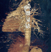
CT reformation of a patient with chronic aortic dissection type A, 25 months following implantation of a hybridprosthesis. The proximal segment of the false lumen covered by the stented segment of the hybridprosthesis is thrombosed.
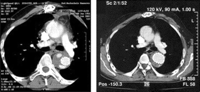
(a,b) Contrast CT-scans of a patient with a chronic aortic dissection type A following implantation of a hybridprosthesis 1 week (left panel) and 10 months (right panel) postoperatively. Perigraft space around the stented segment of the hybridprosthesis in the descending aorta is thrombosed partially at 1 week postoperatively (left panel). At 10 months postoperatively the perigraft space is thrombosed completely. The diameter of the stented segment of the hybridprosthesis became wider when compared to 1 week postoperatively after complete unfolding of the stent. Compared to the status 1 week postoperatively, the diameter of the aneurysm in the descending aorta is now reduced.
Both phenomena, thrombus formation of the perigraft space and shrinkage of the excluded aneurysms, are indicative of a reduction of aortic wall stress thereby reducing the risk of rupture of the stented aortic segment. This finding together with a relatively low perioperative mortality and morbidity supports the therapeutic concept of a single-stage antegrade combined open and endovascular repair of complex thoracic aortic aneurysms using a hybridprosthesis. Further evaluation of this treatment modality appears therefore warranted.
Appendix A
Conference discussion
Dr M. Krason (Zabrze, Poland): I would like to ask you a question and make a remark. Putting the leader down the femoral arteries is much more difficult in this case. We have done several procedures like this with a commercially available Medtronic device and it was easier and better to recognize the true lumen from antegrade through open distal anastomosis. Second, and it's also our problem, and this is the real question, do you feel that partial clotting of the descending thoracic aorta is a good enough solution for these patients?
Dr Karck: No, definitely not. Partial thrombus formation should not be the goal. We are looking for complete thrombus formation, which we in fact have observed in the majority of cases after the follow-up of 14 months. Even after this time, and this is what we know from the conventional stent experience, we may hope that thrombus formation will go on in patients, who have only partial thrombosis formation in the perigraft space so far.
With regard to your remark that antegrade placement is better than retrograde placement, I have to tell you that we had one misplacement of the hybridprosthesis because we did not use the transfemoral through-and-through guidewire system, which we have employed ever since. So, we strongly believe that we should have a guidewire which is forwarded transfemorally either one hour before or directly with the beginning of surgery to be able to properly position the stented segment in the true lumen. You also have to consider the tortuosity of the descending aorta which may put you in trouble if you don't have a guide to guide the introduction of the introducer system.
References
discussion 1891–4
Author notes
Presented at the joint 18th Annual Meeting of the European Association for Cardio-thoracic Surgery and the 12th Annual Meeting of the European Society of Thoracic Surgeons, Leipzig, Germany, September 12–15, 2004.
