


Abstract
Patients who underwent isolated aortic valve replacement could come to attention for new onset aortic disease or progression of borderline alterations not corrected at the first operation, especially in the subset of bicuspid valve disease. We describe our technique in redo operations for aortic root disease, using only a vascular graft and sparing the previously implanted valve prosthesis. In case of normally functioning mechanical prosthesis, we always left the valve in situ and substituted the aortic root with a Dacron conduit, extending the replacement if necessary to the other diseased portions of the thoracic aorta.
1 Introduction
Patients who had previous cardiac operations are an enlarging proportion of the nowadays surgical case mix. Patients who underwent isolated aortic valve replacement (AVR) could come to attention for new onset aortic disease or progression of borderline alterations not corrected at the first operation. Aortic root pathology requiring redo surgery ranges from aneurysms to complications of AVR surgery (pseudo-aneurysm, fistula). Moreover, the subset of patients with bicuspid valve disease is particularly at risk of developing aortic dilatation or dissection [1–3]. Recently, some evidences might prompt to a more aggressive approach of root replacement at time of aortic valve surgery [4], but there is not yet a clear cut-off value of aortic diameters to opt for a conservative management or not. For this reason, i.e. inability to predict aortic disease progression, isolated AVR is often justified and knowledge of different redo surgical approaches are required.
Herein, we describe how to face these kind of situations using only a vascular graft and sparing the previously implanted valve prosthesis. In case of normally functioning mechanical prosthesis, we left the valve in situ and substituted only the aortic root, if necessary extending the replacement to other diseased portions of the thoracic aorta.
2 Technique
The surgical approach included repeat sternotomy (Fig. 1, panels A). Femoral artery, aortic arch or axillary artery were used as arterial inflow. The aorta was fully mobilized in cardiopulmonary bypass. After cardioplegic arrest, aorta was dissected from the other structures, the aneurysm was excised and the coronary isolation was then completed.
The prosthesis was carefully inspected for thrombi, pannus, or reduced mobility of the leaflets. The base of the aorta was then trimmed to leave a 4mm remnant and the Dacron conduit size was definitely chosen (Fig. 1, panels B). We are now currently implanting a modified tube graft that incorporates pseudosinuses of Valsalva because it seems to facilitate anatomical reconstruction of the aortic root and it has the potential to positively affect the coronary flow [5]. Horizontal Teflon felt-pledgeted 2–0 mattress sutures were placed from outside the aortic annulus to the sewing ring of the previously implanted valve and then to the conduit (Fig. 2, panels C). The graft was parachuted and sutures knotted. Tube/left ventricle outflow tract coupling can be tested by forced instillation of saline, the valve closed and the graft obliquely clamped. A running suture between the cut edge of the aortic wall and the proximal portion of the tube further improves hemostasis [6]. The placement of the orifice for the right coronary artery was chosen after a short clamping of the venous line, allowing for right ventricle dilatation. Each coronary ostium was anastomosed by a simple running suture or by an endobutton-buttress suture, i.e. incorporating peri-ostial aortic tissue. Anastomoses were tested by infusion in the graft of cardioplegic solution or saline. The distal graft is finally trimmed according to the anatomy of the distal aorta or to the arch prosthesis (Fig. 2, panels D). Prosthetic valve endocarditis is a clear indication to remove the valve whereas paraprosthetic leak can be repaired by the same pledgeted sutures.
3 Comment
We adopted the present strategy in 4 patients. All procedures were completed and no conversion to composite graft recorded. Operative times were shortened compared to the implantation of composite graft obtaining a potential benefit for a faster recovery. Bleeding from the proximal suture was never matter of revision.
The described technique avoids implantation of a composite graft on a weakened or traumatised aortic annulus, after removal of the previous prosthesis, and it may reduce the risk of pseudo-aneurysm formation. Moreover, use of a shaped Dacron graft seemed to facilitate coronary button reimplantation. Actually, in redo operations, the coronary arteries are difficult to mobilize and remain frequently displaced. This kind of prosthesis reduces the distance from the coronary ostia and may decrease tension upon the sutures. No interposition material was required in our experience.
Improved surgical techniques and perioperative management have reduced morbidity and mortality associated with aortic valve surgery allowing for a prolonged expectance of life. In this context, our patients could develop aortic disease independently from the valve pathology, but progressive aneurismal dilatation or aortic dissection are more and more linked to the underling aortic valve disease [1,7] or to systemic syndromes [8]. Only in Marfan patients, values of aortic diameter have been defined to prompt prophylactic aortic replacement; in other clinical conditions is still difficult to establish the need of aortic root substitution at surgery for the aortic valve. For these reasons, redo operation after AVR is a probable clinical scenario. Therefore, mastering different techniques in aortic root surgery, like the described one, might be useful to proceed electively with root replacement.
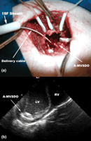
(a) The A-MVSDO completely opened connected with delivery cable through the substernal incision (18F sheath—external diameter 8mm). (b) IVUS echocardiogram showing the A-MVSDO completely opened on the LV wall.
Dr Emmanuel Villa was the Traveling Fellow 2003 of the Italian Society for Cardiac Surgery.
