-
PDF
- Split View
-
Views
-
Cite
Cite
Ayten Kayı Cangır, Cabir Yüksel, Mehmet Dakak, Enver Özgencil, Onur Genc, Hadi Akay, Use of intrapleural streptokinase in experimental minimal clotted hemothorax, European Journal of Cardio-Thoracic Surgery, Volume 27, Issue 4, April 2005, Pages 667–670, https://doi.org/10.1016/j.ejcts.2004.12.043
Close - Share Icon Share
Abstract
Objective: In clotted hemothorax, both thoracocentesis and closed tube thoracostomy will not be able to evacuate the pleural cavity especially if it is minimal. The aim of this study was to assess the effectiveness of intrapleural administered streptokinase on minimal clotted hemothorax without drainage, in order to accelerate the spontaneous resolution and absorption in blunt thoracic trauma. Methods: Thirteen adult ewes were used for this experiment. The animals were divided into two groups. First group served as the control group (Group C) (n=5) and did not receive any intrapleural fibrinolytic treatment. In both groups, 200ml of blood was taken from the left jugular vein and injected into the pleural cavity with a serum line through the scope after pleural abrasion. Streptokinase (150.000U) was diluted in 100ml of saline and applied to the second group (Group S) (n=5) in second postoperative day. One ewe in each group was sacrificed with a lethal dose of sodium thiopental in postoperative 2nd, 4th, 6th, 8th, and 10th weeks, respectively. When a left posterolateral thoracotomy was performed, pleural thickening and adhesion were evaluated. The lung and pleural tissue samples were taken for histopathologic examination. The slides were examined in a blinded manner. Results: Thoracentesis was performed in all ewes in the second postoperative day and no fluid was detected. There was no allergic reaction in group S after the injection of streptokinase into the pleural cavity. During postmortem macroscopic evaluation, we observed clot in one of the ewes in group C in second postoperative week. A statistically significant difference was found between Group C and S regarding pleural thickening and adhesion (P=0.05). The ewes of Group S had less pleural thickening and adhesion compared to those of Group C. These results were confirmed with histopathological examination. Conclusion: We conclude that intrapleural streptokinase increases resolution of clot in the pleural space and decreases pleural thickening and adhesion in experimental minimal clotted hemothorax in ewes. This study has also demonstrated that intrapleural streptokinase can be used without drainage. Use of intrapleural streptokinase without drainage can be a novel therapeutic option for trauma patients with minimal clotted hemothorax after haemorrhage of other organs was excluded.
1 Introduction
Management of hemothorax is an important problem in thoracic surgery. The traditional initial treatment of traumatic hemothorax is closed tube thoracostomy drainage. However, closed tube thoracostomy drainage is ineffective in clotted hemothorax. Insufficient treatment carries a number of problems in posttraumatic clotted hemothorax [1–3]. Hemothorax may become infected if a pneumonic process or a persistent bronchopleural fistula or major sources of sepsis elsewhere in the body occur. Besides, repeated thoracentesis is an additional risk for infection. In untreated hemothorax, the pleural surface is covered by a thin layer of fibrin and cellular elements. Angioblastic and fibroblastic proliferation occurs after seven days [4]. Also a continuous thickening of the membrane by progressive deposition and organization of the coagulum within the pleural cavity occur. These cases may require decortication to mobilize and expand the lung with thoracotomy or video-assisted thoracoscopic surgery (VATS) [2,3,5]. However, a certain number of clotted hemothoraces resolve spontaneously. Thoracotomy and VATS are invasive procedures and are associated with potential morbidity especially in patients with trauma. In addition, some authors suggest the fibrinolytic treatment through the chest tube as a treatment modality in traumatic clotted hemothoraces [1,6].
In this study, we have investigated the role of intrapleural administered streptokinase (SK) on minimal clotted hemothorax without drainage, in order to accelerate spontaneous resolution and absorption in blunt thoracic trauma.
2 Materials and methods
Thirteen adult ewes were used for this experiment. Animal acquisition was under the supervision of the Gülhane Military Medical Academy Animal Research Laboratory and a licensed veterinarian. All animals involved in this study received animal care in compliance with ‘Principles of Laboratory Animal Care’ formulated by the National Society for Medical Research and the ‘Guide for the Care and Use of Laboratory Animals’ published by the ‘National Institutes of Health’ (NIH publication 85–23, revised 1985). This study was approved by the relevant institutional ethic committees.
The animals were divided into two groups. First group served as the control group (Group C) (n=5; body weight, 35–42kg) and did not receive intrapleural fibrinolytic treatment. Intrapleural SK was applied to the second group (Group S) (n=5; body weight, 35–42kg) in second postoperative day.
2.1 Operative model
All animals were premedicated with 0.5mg/kg intramuscular atropine sulphate. At the same time, 1mg/kg intramuscular ketamine was used for anesthesia induction. Approximately half-hour after induction of anesthesia, endotracheal tube was inserted and continuous anesthesia was provided with 1–2% halothane in 50% O2+50% air mix. A catheter was inserted into the left jugular vein. Animals were placed in the usual full left lateral thoracotomy position and then a port was placed in the midaxillary line of the sixth or seventh intercostal space. The scope was introduced and all organs in the left hemithorax were explored (Fig. 1a). A hydatid cyst was observed in an animal and it was excluded from the study. After the exploration, pleural abrasion was performed using a peanut at the parietal pleura. Then, 200ml of blood was taken from the jugular vein and injected into the pleural cavity (Fig. 1b). The port was closed in layers during positive ventilation. All animals were extubated after the spontaneous respiration had begun. One ewe died because of respiratory insufficiency. Each animal received prophylactic antibiotic (Cefazoline) and 500ml 0.9% NaCl. In the second postoperative day, thoracentesis was performed in all animals in order to control the presence of clotted hemothorax. In group S, fibrinolytic agent (SK 150.000U) mixed with 100ml of normal saline was injected through the intercostal space one below via 21 gauge needle after the second postoperative day.
One ewe in each group was sacrificed with a lethal dose of sodium thiopental in postoperative 2nd, 4th, 6th, 8th, and 10th weeks, respectively. When a left posterolateral thoracotomy was performed, pleural thickening and adhesion were evaluated according to changes in Table 1.
After macroscopic evaluation, lung and pleural tissue samples were taken for histopathologic examination. The slides were examined in a blinded manner. In histopathologic evaluation, one of the ewes in the control group had the diagnosis of adenocarcinoma so this ewe was excluded from the study.
The distribution of these variables was compared within the groups. The univariate statistical analysis was done by Mann–Whitney U-test. Analyses were performed using SPSS for Windows 10/0 (SPSS, Inc., Chicago, IL). Differences were considered as statistically significant, when the P value was less than 0.05 or equal.
3 Results
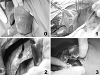
Thoracentesis was performed in all ewes in the second postoperative day and no fluid was detected. There was no allergic reaction in group S after the injection of SK into the pleural cavity. Postmortem macroscopic evaluation was done according to the scoring in Table 1 and findings were shown in Table 2 (Fig. 2). During postmortem macroscopic evaluation we observed clot in one of the ewes in group C in second postoperative week. Severe pleural thickening and firm pleural adhesions were not observed in any groups. A statistically significant difference was found between Group C and S regarding pleural thickening and adhesions (P=0.05). The ewes of Group S had fewer pleural thickening and adhesion compared to those of Group C. Results of postmortem macroscopic evaluation was confirmed with histopathological examination.
4 Discussion
Hemothorax commonly occurs following blunt chest trauma. Major operations are rarely required. There is a general agreement that the initial treatment of patients with traumatic hemothorax includes chest tube drainage [4,7]. However, when hemothorax is minimal, chest tube drainage may not be required and these cases can be followed. Besides, after blunt chest trauma, intrapleural blood may coagulate and chest tube drainage may not be possible especially if the clotted hemothorax is minimal [4]. Treatment of the minimal and/or clotted hemothorax remains controversial.
Computed tomography (CT) scan has been used for evaluation of chest traumas for three decades. More detailed findings were obtained with CT scan than with X-ray or physical examination as it is difficult to detect presence of minimal hemothorax by those methods. Therefore diagnosis of hemothorax is easily made by CT scan even it is minimal. The number of cases diagnosed as minimal hemothorax after chest traumas increased with the routine usage of CT scan.
Intrapleural blood almost universally clots in the early post injury period [4]. A thin covering of the pleural surface by fibrin and cellular element occurs. This covering develops into a progressively thicker membrane that coats the visceral and parietal surface and forms a saclike structure containing the hemothorax [4]. The clotted hemothorax should be evacuated within 7–10 days of injury to prevent the formation of fibrous peel and to reduce the risk of empyema. Therefore some authors suggest that intrapleural fibrinolytic treatment or VATS can be performed in treatment of clotted hemothorax [1–3,5]. However, management of minimal hemothorax to prevent from pleural thickening without chest tube insertion is not clear. Condon et al. [8] reported that pleural blood may be spontaneously absorbed. However, clot cannot be absorbed spontaneously, and will even cause respiratory embarrassment, become infected, or evolve to fibrothorax, and finally require decortication [5,9]. Regarding these complications, treatment of clotted hemothorax is required even it is minimal. Besides, after 2–4 weeks elapse, the clot begins to lyse and this results in a markedly hypertonic pleural collection. The body responds to this by rapidly secreting a large quantity of fluid in the pleura and a thoracentesis or an intercostal tube insertion may be necessary. We thought that this process can be facilitated by intrathoracic fibrinolytic treatment in minimal hemothorax without an intercostal tube insertion. We designed this experimental study before performing this treatment method in trauma patients. Therefore we evaluated the efficacy and safety of intrapleural administration of 150.000U SK to enhance the resolution of experimental minimal hemothorax in the ewes' pleural spaces.
Complications of the therapeutic use of intrapleural fibrinolytic agents are not frequent. Intrapleural SK instillation may cause coagulopathies or allergic reactions. Besides, side effects appear to be less frequent with a putrefied form of SK. We found only one experimental animal study regarding the use of intrapleural SK in the literature [10]. Strange and colleagues reported that intrapleural SK has been used in multiloculated empyema in the rabbit's pleural space. In our study no allergic reaction or coagulopathy were observed after injection of SK into the pleural space in group S, similar to the study of Strange et al. [10].
Streptokinase degrades a variety of proteins, including fibrin. Therefore, intrapleural SK is useful in the treatment of persistent, loculated hemothorax and empyema [6,7]. Administration of intrapleural SK avoids surgical decortication in a significant number of patients as other aggressive, prolonged, and expensive therapeutic options. The goal of the treatment in clotted hemothorax is rapid removal of clot before formation of pleural thickening and adhesion. Some authors recommended VATS in the management of patients with retained hemothoraces to avoid the problems of secondary infection within the intrathoracic clot or late fibrothorax [2,11]. Similar problems can occur in patients with minimal clotted hemothorax.
Acute bleeding might occur using intrapleural fibrinolytic treatment in traumatic patients, therefore SK was injected after the second postoperative day in this study. In clinical practise, after excluding the haemorrhage of other organs, massive hemothorax, acute bleeding or bronchopleural fistula, SK may be injected into the pleural cavity of traumatic patients in the second day after trauma.
In this study, we compared the pleural thickening and adhesion of two groups, and found that the findings in group C was worse than the ones in group S and this difference was statistically significant (P=0.05). We thought that use of SK accelerated the lysing of post traumatic intrapleural clot and the period of absorption became shorter. Therefore fewer pleural thickening and adhesions were occurred in group S.
We conclude that intrapleural SK increases resolution of clot in the pleural space and decreases pleural thickening and adhesion in experimental minimal clotted hemothorax in ewes. This study has also demonstrated that intrapleural SK can be used without drainage. Use of intrapleural SK without drainage can be a novel therapeutic option for trauma patients with minimal clotted hemothorax after the haemorrhage of other organs, massive hemothorax, acute bleeding or bronchopleural fistula was excluded. Clinical investigations about the use of SK in trauma patients with minimal hemothorax are needed for further conclusions.

(a) A port was placed in the midaxillary line of the sixth or seventh intercostal space, the scope was introduced and all organs in the left hemithorax were explored, (b) 200ml of blood was taken from the jugular vein and injected into the pleural cavity with a serum line through the scope after pleural abrasion.

Scoring of pleural thickening and adhesion; (0) no change in visceral or parietal pleura, (1) mild pleural thickening or loose pleural adhesion, (2) moderate pleural thickening or firm pleural adhesion, (3) severe pleural thickening or firm pleural adhesion.


References
- streptokinase
- fibrinolytic agents
- chest injury trauma blunt
- process of absorption
- hemorrhage
- lung
- hypersensitivity
- adhesions
- adult
- autopsy
- hemothorax
- thiopental
- wounds and injuries
- pleura
- sodium
- thoracentesis
- pleural space
- normal saline
- tube thoracostomy
- abrasion
- posterolateral thoracotomy
- pleural fibrosis
- lethal dose
- histopathology tests


