


Abstract
Objective: Although several methods of stent-grafting for patients with aortic arch aneurysm have been reported, these methods have been associated with several complications such as endoleak and migration. We developed a new method using Matsui-Kitamura (MK) stent-graft following extra-anatomic arch vessels bypass by selective cerebral perfusion (SCP) under left heart bypass (LHB). Methods: Between December 2001 and December 2003, 12 patients with aortic arch aneurysm were treated according to this new method. All patients were male with an average age of 71.3 ± 6.4 years. There were 5 patients with severe pulmonary dysfunction, 4 with renal dysfunction, one with severe cardiac dysfunction and 3 with preoperative cerebral infarction. Under SCP using LHB, the extra-anatomic arch vessel bypass was established. The MK stent-graft was delivered into the aortic arch. Coronary artery bypass grafting (CABG) was concomitantly performed in one patient. Results: There were no cases of endoleak, migration or hospital death. One patient, who had a past history of cerebrovascular disease, suffered a minor stroke, and one patient, who was performed CABG to the mid-left anterior descending branch (LAD) using the left internal thoracic artery (LITA), presented paraparesis. Although two patients of chronic renal failure underwent scheduled CHDF on account of using the contrast medium during the procedure, all of them were weaned from hemodialysis. However, there were no other postoperative complications such as, respiratory failure or cardiac dysfunction. Conclusions: Endovascular stent grafting EVSG using the MK stent with extra-anatomic arch vessel bypass under SCP using LHB could be a useful and less invasive method for patients with aortic arch aneurysm who are at a high surgical risk.
1 Introduction
Conventional surgical therapy for a thoracic aortic aneurysm is getting better results thanks to recent advances regarding the surgical procedure [1,2]. However, patients with renal dysfunction, respiratory dysfunction or of an old age still are at a very high risk of considerable morbidity and mortality [3]. Endovascular stent grafting is less invasive than conventional aortic surgery for aortic aneurysm, and recently, its use has been extended to aortic arch aneurysm [4]. Although this is a less invasive procedure and a new option to treat patients with thoracic aortic aneurysm, stent-grafting for thoracic aortic aneurysm, especially for aortic arch aneurysm, presents technical and anatomical difficulties. A proximal landing zone of at least 2cm is required for endovascular stent grafting. However, the cervical branches that originate from the aortic arch are an obstacle to obtain a sufficient landing zone. Thus there have been many reports describing aortic arch reconstruction using an extra-anatomical bypass [5–7]. Another problem is that aortic arch is severely curved. Using a rigid type of stent-graft constructed with a Z stent and graft might not be tightly fitted to the curvature of the aortic arch. The Matsui-Kitamura (MK) stent-graft (Kitamura Inc., Kanazawa, Japan) was designed to fit the curvy segments of the aorta [8]. And we developed a new stent-grafting technique using the MK stent-graft following bypass grafting of arch vessels under left heart bypass to prepare the landing zone for the stent-graft [9]. We applied successfully the new method in 12 patients with aortic arch aneurysm.
2 Materials and methods
Between December 2001 and December 2003, 12 patients with aortic arch aneurysm were treated according to the new method using the MK stent-graft. All patients were male, and the average age of the patients was 71.3±6.4 years old (range, 52–76). All patients in this series presented severe comorbid medical conditions. Average Euro SCORE of the patients was 6.4±1.8. There were 5 patients with severe pulmonary dysfunction, 4 with renal dysfunction, one with severe cardiac dysfunction and 3 with preoperative cerebral infarction (Table 1).
Preoperative evaluation of the entire aorta was done by contrast-enhanced multi-detector row helical computed tomography (Aquilion; Toshiba Medical Co., Tokyo, Japan). The diameter of the stent-graft was determined on the basis of the CT findings. Multi-planar reconstruction and three-dimensional imaging were very useful to demonstrate the morphological details of the aneurysm and femoral artery from where the sheath system of the stent-graft was approached. A diameter 20% greater than that of the ascending aorta was selected as the diameter of the MK stent-graft.
The MK stent-graft was constructed from a framework of MK stent and a polyester fabric graft. The framework of the MK stent is weaved with a single length of NiTi wire with a diameter of 0.4mm. NiTi alloy was used to shape a metallic stent that consisted of 51wt% of nickel and 49wt% of titanium (Memoalloy, Tokin Inc., Tokyo, Japan). The wire was wound and connected both ends of the wire with a compressed, small platinum cylinder. The diameter and length of the stent-graft were determined for each patient from measurements obtained from preoperative diagnostic images. The curved MK stent was covered with a seamless, cylindrical graft made of a thin polyester fabric of 0.1mm in thickness. The graft was attached to the framework of the stent at both ends with a 5-0 monofilament suture, since full covering might prevent full expansion of the stent. The MK stent-graft could be deployed through an 18 Fr or 22 Fr J-shaped sheath system (Cook Ind., Bloomington, IN, USA).
The operative procedure was typically performed through a full sternotomy. Prior to full sternotomy, bilateral subclavian arteries and the right femoral artery were exposed and taped. At the same time, a straight conduit was anastomosed to a side of a Dacron Y graft (proximal diameter, 16mm) to triplicate the outflow tract of the graft. After full sternotomy, LHB was established with cannulation of the right upper pulmonary vein for inflow and cannulations of bilateral subclavian arteries for outflow after general heparinization, and the activated coagulation time (ACT) was kept at about 200s. LHB was switched to antegrade SCP by placing clamps on 3 arch vessels and inserting an additional arterial cannula into the left common carotid artery. A roller pump was used for right subclavian artery perfusion, and a centrifugal pump was used for perfusion of the left subclavian artery and the left common carotid artery. Flow rates of the each pump were maintained at 10mL/kg per minutes. Perfusion pressure was monitored by the perfusion catheters of the right subclavian artery and the left common carotid artery, and was adjusted to maintain more than 60mmHg. During the period of antegrade SCP, patients were kept at mild hypothermia (33°C bladder temperature). The arch vessels were divided, and their stumps were closed with 5-0 monofilament sutures. A side-biting clamp was placed on the ascending aorta, and the proximal end of the Y graft was anastomosed to the aorta in an end-to-side fashion using 5-0 monofilament sutures. Subsequently, the side-biting clamp was removed, and the three outflow tracts of the Y graft were anastomosed to the 3 arch vessels, respectively, in an end-to-end fashion. After weaning from SCP, protamine was administered once to achieve hemostasis. A sufficient landing zone was prepared for the proximal side of the stent graft by means of this extra-anatomical bypass. After general reheparinization, and the ACT was kept at about 200s, a 5-Fr sheath (Terumo Co., Tokyo, Japan) was inserted into the ascending aorta, an 8-Fr sheath (Terumo Co.) was inserted into the right femoral artery, a 0.032-inch guide wire (Terumo Co.) was inserted into the aorta through the 5 Fr sheath, and the wire was pulled out from the sheath in the right femoral artery using a snare catheter. A long sheath catheter as stent-graft delivery system was advanced into the target segment of the aorta through a transverse arteriotomy of the right femoral artery over the guidewire using tug of wire methods [10]. The custom made MK stent graft was delivered by a long sheath catheter. Then a 5-Fr angiographic catheter was placed into the ascending aorta. Aortography was performed using 20ml of contrast medium to confirm the relationship between the aneurysm and the proximal end of the Y graft anastomosing site of the ascending aorta. After the aortography, the stent-graft was deployed in the correct position. Immediately after deployment, post-dilation was achieved with a TMP lock balloon catheter (Tokai Medical Product Co., Aichi, Japan) (Fig. 1). Aortography was performed again to make sure that there was no endoleak and migration of the stent-graft.
3 Results
Endovascular placement of the curved MK stent-graft was successful without endoleak or migration in all patients. None of the patients died in hospital and 30 days after the operation, the mortality rate was 0%. The diameter of the MK stent-graft was 39±2.28mm (range, 34–42mm). One patient, who had a history of cerebrovascular disease, suffered with a minor stroke. After 2 months in a rehabilitation program, he almost fully recovered from his left side paralysis. One patient, who was performed CABG to the mid-LAD using LITA, presented paraparesis. This patient was able to walk with a stick after rehabilitation training. There was no other postoperative complication such as renal dysfunction requiring hemodialysis, respiratory failure requiring prolonged ventilator support (≫72h) or cardiac dysfunction. The mean follow-up was 15.7 months (range, 4–29 months) for 10 patients. Nine patients could be followed up with CT examination every six months after the operation. Neither late endoleak, migration, aneurysm diameter enlargement, nor aneurysm rupture was observed. One patient died of unknown reasons 6 months postoperatively. In all nine patients, the aneurysmal space was completely thrombosed on CT examination, and six of nine patients showed reduction of aneurysm in size (Fig. 2). The other three patients have not shown a change in the size of the aneurysm (Table 2).
4 Discussion
Various approaches and extracorporeal circulation technique have been described for the surgical treatment of aortic arch and distal aortic arch aneurysm. Recently, EVSG of aortic arch aneurysm for high-risk patients, who present complications such as cerebrovascular accident, respiratory dysfunction, or renal dysfunction, was described. Although EVSG is minimally invasive, initial success rate of EVSG for aortic arch aneurysm was not too good because of the anatomical characteristics of the aortic arch.
One of the reasons for the difficulty of EVSG therapy for patients with aortic arch aneurysm is that there is only a short proximal landing zone, resulting in a high rate of proximal endoleak. In general, a landing zone of at least 2cm is necessary for a standard stent-grafting technique because tight in that way a fixation of the stent-graft to the aortic wall is achieved without endoleak. Almost all patients with aortic arch aneurysm needed relocation of the branching vessels to obtain a sufficient landing zone. Recently, the several methods to create a longer proximal landing zone using a combined surgical procedure, were reported [5–7]. In most reports, relocation of the branching vessel was performed by extra anatomical bypass under simple clamp without any circulatory support. On the other hand, some reports concerning this method have described a significant occurrence of neurologic complications and a high mortality rate [11]. The neurologic complications in this method may result from an interruption of the cerebral circulation caused by embolization of atherosclerotic debris. We applied antegrade SCP using LHB to relocate of the branching vessels, so that cerebral perfusion was not interrupted. Kazui et al. reported a low frequency of neurologic complications in patients subjected to antegrade SCP [1,12]. When a side-biting clamp was placed on the ascending aorta to anastomose the proximal end of the Y graft to the aorta, antegrade SCP was already started and the proximal end of the branching vessels were clamped. Thus, neurologic complication due to atherosclerotic debris from the wall of the ascending aorta was prevented. Moreover, when we carry out EVSG, neurologic complications due to catheter manipulation in the aortic arch are also a serious problem. In our technique, all the arch vessels have already been bypassed, when we manipulated the catheter in the aortic arch. Thus, it is safer than the conventional EVSG technique. Besides the advantages of endovascular stent graft delivery with extra-anatomic arch vessel bypass by SCP are that with this technique we can avoid cardiac arrest, circulatory arrest and the use of an oxygenator. Our results showed that there was no postoperative complication such as renal dysfunction requiring hemodialysis, respiratory failure or cardiac dysfunction, even in patients considered to be at a high surgical risk. This suggested that the surgical stress inherent to this procedure was minimum and acceptable for patients. SCP requires a lower level of heparin as compared with cardiopulmonary bypass because an oxygenator is not used. It is a big advantage of this procedure to prevent bleeding in a patient with such potential for massive pulmonary bleeding.
Another reason for the difficulty to perform EVSG for patients with aortic arch aneurysm is the severely curved configuration of the aortic arch. A rigid, straight, tube-type stent-graft suffers late deformation or migration causing thereby an endoleak or aortic wall injury [13]. The proximal portion of a stent-graft may cause pulsatile stress on the native aortic wall and may injure the wall, because of the mismatched configuration between the stent-graft and the aortic arch. Particularly, in a patient with a short proximal neck, the proximal anchoring bare segment of the stent-graft is placed across the orifice of the left subclavian artery, and may directly injure the aortic wall or the orifice of the subclavian artery. The curved configuration of the MK stent accommodated to the aortic arch and thus a late deformation or migration of the stent-graft was reduced, preventing thereby the occurrence of an endoleak and minimizing injury to the aortic wall. The MK stent graft is a flexible, custom-made, curved stent graft. In these cases the MK stent graft fit tightly to the 3-dimensional curvy portion of the aorta and did not cause kinking or endoleak at all. Although total replacement of the aortic arch would be one of the best solutions for an aortic arch aneurysm, this procedure is surgically less stressful, especially for patients with preoperative complications.
5 Conclusion
New stent-grafting using an MK stent-graft following bypass grafting of arch vessels under left heart bypass might be useful for patients with aortic arch aneurysm complicated by severe organ dysfunction or for aged patients.
Appendix A. Conference discussion
Dr T. Carrel (Bern, Switzerland): Why do you use cardiopulmonary bypass? We have done a similar operation without cardiopulmonary bypass. You can first debranch every supra-aortic vessel, and then once you have started reperfusion on the brain and the arms, you can easily introduce the stent either antegradely through the arch or through the ascending or in a retrograde fashion. Using this technique you may prevent all negative effects of cardiopulmonary bypass and, of course, of hypothermic circulatory arrest and cerebral perfusion.
Dr Akasaka: Some papers say that if the cervical branches are grafted with temporary bypass there is a very high percentage or incidence of cerebral accident. So we prepared against cerebral accident. We used a left heart bypass to graft the arch vessels.
Dr Carrel: But I do not see the reason to use left heart bypass. Especially if you have an atherosclerotic aortic arch, you might dislodge thrombus or atherosclerotic material through manipulation during cannulation for instance. So it might be better to first debranch every supra-aortic vessel, to close them, and then to manipulate into the arch after you have perfusion of the brain through the grafts from the ascending aorta.
Dr Akasaka: We applied the side-clamp to the ascending aorta and selective cerebral perfusion was started and the arch vessels were already clamped. Therefore we can prevent the embolization of the cerebral artery, which is caused by the side-clamp of the ascending aorta. However, we must cannulate the arch vessels to establish selective cerebral perfusion. We could not prevent cerebral infarction due to cannulation of the arch vessels. So I think we have some risk of cerebral infarction in the new method, but the selective cerebral perfusion time in our method was very short compared with conventional arch replacement. So I think our method is still really impressive concerning cerebral protection, as our results show that we have only one patient who had a complicated cerebral infarction, though three patients had cerebral infarction in their past history. So I think our method is still safer.
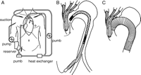
(A) The assisted circulation is shown. Left heart bypass was switched to SCP by placing clamps on 3 arch vessels. (B) After the arch vessels were divided and closed, the extra-anatomical bypass was constructed. A stent-graft delivery system was advanced into the aortic arch. (C) An extended aortic arch aneurysm was treated with the MK stent graft, which was constructed from a nitinol framework of MK stent and polyester fabric and inserted into the aortic arch.
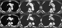
A 67-year-old male with aortic arch aneurysm. (A) Preoperative enhanced CT scan demonstrates a true sacclar aneurysm in the aortic arch. (B) Enhanced CT scan obtained 1 month after MK stent-graft placement revealed complete exclusion of the aneurysm without endoleak. (C) 9 months after the procedure, enhanced CT scan showed that the aneurysm in the aortic arch was almost vanished.
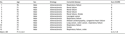
Characteristics of the 12 patients with aortic arch aneurysms treated with the new method using the Matsui-Kitamura stent-graft following extra-anatomic arch vessels bypass by selective cerebral perfusion under left heart bypass
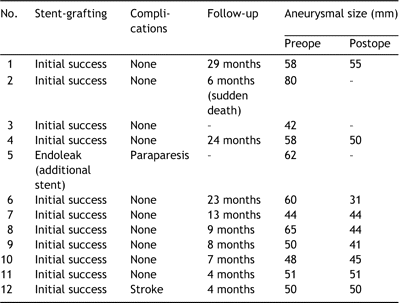
Results of the new method using Matsui-Kitamura stent-graft
Presented at the joint 18th Annual Meeting of the European Association for Cardio-thoracic Surgery and the 12th Annual Meeting of the European Society of Thoracic Surgeons, Leipzig, Germany, September 12–15, 2004.
