-
PDF
- Split View
-
Views
-
Cite
Cite
Shunsuke Endo, Yuji Sakuma, Yukio Sato, Yasunori Sohara, Unusual chronic rheumatoid empyema, presenting as a chest wall mass, European Journal of Cardio-Thoracic Surgery, Volume 26, Issue 5, November 2004, Page 1042, https://doi.org/10.1016/j.ejcts.2004.08.001
Close - Share Icon Share
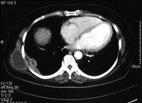
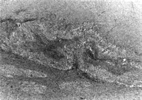
A 50-year-old female with rheumatoid arthritis who had a right pleuritis 2 years earlier, had a chest wall mass as shown in Fig. 1. Erythrocyte sedimentation rate was 48mm/h. A cystectomy was performed. Pathological study indicated a necrobiotic cyst containing rheumatoid nodules (Fig. 2). Neither fungus nor bacteria were identified.

Enhanced chest computed tomography shows a protrusive chest wall mass measuring 5×5×3cm, communicating into the right pleural thickening.

Pathologic findings shows rheumatoid nodule containing layer of palisaded epitheloid histiocytes adjacent to necrotic debris.


