


Abstract
Objectives: Although endovascular stent-grafting is available for atherosclerotic thoracic aneurysms, it is unknown whether the excluded thrombosed aneurysms shrink. We evaluated serial changes in distal aortic arch or proximal descending aortic aneurysms excluded space after transaortic stent-grafting. Methods: Thirty-four patients with true distal aortic arch or proximal descending thoracic aortic aneurysms were treated by stent-grafts introduced via proximal arch aortic incisions. Follow-up included computed tomographs (CT) every 6 months in 31 patients. The maximum dimensions for excluded space and aneurysmal diameters were measured and evaluated to determine whether the aneurysmal space had decreased or shrunken following this alternative procedure. Results: Two hospital deaths (5.9%) were caused by a cerebral embolism and a peri-operative myocardial infarction. Another case died from pneumonia a year after surgery. Thirty-one cases (91%) survived during follow-up, but one case suffered from paraplegia (2.9%). The follow-up period ranged from 10 to 72 months (average 39.3±27.2 months). There were no aneurysmal ruptures during follow-up. Post-operative serial CTs showed a disappearance or a significant shrinkage of the excluded aneurysmal space in 30 of the 31 cases (97%); one case suffered endoluminal leakage. Conclusions: Transaortic endovascular stent-grafting is feasible for distal aortic arch or proximal descending aortic aneurysms. The excluded aneurysmal space disappears or shrinks after successful stent-graft placement.
1 Introduction
There are two different procedures for endoluminal repair of thoracic aortic aneurysms. One is transcatheter endoluminal stent-graft placement [1,2]. Transcatheter stent-graft placement is an innovative technique, especially in the treatment of descending thoracic aortic aneurysms [3]. The other procedure, transaortic stent-grafting, is used in distal arch or proximal descending aortic aneurysms [4,5]. The percutaneous insertion of the stent-graft is often difficult to adapt to the branched and curved aortic arch; in this type of aneurysm, the proximal neck cannot be sufficiently secured. Since a distal aortic arch aneurysm is often situated in the root of the left subclavian artery, endoluminal aneurysmal coverage may cause left subclavian artery obstruction or endoluminal aneurysm leakage because of inadequate stent-graft placement. Kato and colleagues devised an endoluminal stent-grafting procedure through a circumferential aortic arch incision [4]. We modified their method, and report a simple procedure [5] in which a self-expandable stent can be inserted into the descending aorta through a small proximal arch aortic incision. We named this procedure transaortic endovascular stent-grafting. In this study, we evaluated the feasibility of this alternative procedure for atherosclerotic distal arch and proximal descending aortic aneurysms by serial CT examinations and investigated the changes to the excluded aneurysmal spaces.
2 Materials and methods
2.1 Patients
Thirty-four patients with true distal aortic arch or proximal descending thoracic aortic aneurysms were enrolled in this study. There were 28 men and six women, and their mean age was 69.9±10.9 years (52–88 years old). All cases had atherosclerotic thoracic aortic aneurysms, located at the root of the left subclavian artery, extending into the descending thoracic aorta. All the patients had received a transaortic stent-graft procedure within the last 6 years. Nine cases had proximal aneurysmal extension above the left subclavian artery, and two had subclavian arterial aneurysms. Maximum aneurysmal diameters ranged from 5.2 to 8.4 cm (mean diameter: 6.6±1.5 cm). Out of 34 cases 12 had other cardiac disorders, including 9 with severe coronary artery disease, 2 with aortic valve regurgitation and 1 with tricuspid valve regurgitation. Five patients had stenosis of the iliac and/or femoral arteries. Concomitant surgeries were performed in 22 (22/34, 65%) cases, including 9 coronary arterial bypasses, 2 aortic valve replacements, 1 tricuspid valve annuloplasty, 5 subclavian–subclavian arterial bypasses and 5 total arch replacements. A subclavian–subclavian arterial bypass was selected in cases with left subclavian arterial aneurysms or aneurysmal extension proximal to the left subclavian artery.
2.2 Procedure for stent-graft insertion
All patients were operated upon via median sternotomy. Hypothermic circulatory arrest and selective cerebral perfusion was conducted at 25 °C rectal temperature by extracorporeal circulation. Arterial cannulation for extracorporeal circulation was accomplished via the right subclavian artery with a small prosthesis (8 mm in diameter, ringed PTFE, Gore-Tex, Minneapolis, MN), or the ascending aorta; there was an exception in two cases of calcified ascending aortas which received retrograde femoral arterial perfusion. The small prosthesis was used as a conduit for subclavian–subclavian arterial bypass after extracorporeal circulation termination. Selective cerebral perfusion was performed by direct cannulation into three cervical branches and continued with a blood flow of 600–900 ml/min during hypothermic circulatory arrest and open anastomosis.
Stent-graft insertion was previously described [5]. A Gianturco Z-stent (30 or 40 mm diameter, 5 cm length; Cook, Inc., Bloomington, IN) was inserted into the distal end of the Dacron graft (porosity 250 ml/m2, with a 22–34 mm diameter; Intervascular, Inc., Clearwater, FL) and sutured to the graft. Multiple sized stent-grafts were pre-operatively prepared and sterilized. Stent-graft size was decided by measuring the intact descending thoracic aortic diameter by intraoperative transesophageal echocardiography (TEE). The stent-graft was introduced into a sheath catheter with a 30 F diameter. The distal end of the stent-graft was adequately positioned under TEE guidance [6] and dilated with a large balloon catheter to attach the stent-graft to the descending aortic wall. The proximal end of the stent-graft was circumferentially sutured onto the intact aortic arch wall. The aortic arch incision was closed by the anterior stent-graft wall (Fig. 1) . Subclavian–subclavian arterial bypass was performed in cases with aneurysmal extension proximal to the left subclavian artery. Most of the cases (29 of 34 cases) were treated as indicated above, and termed as conventional transaortic stent-grafting. In five cases with large aneurysms that extended into the proximal and distal aorta, the aortic arch was transected proximal to the left subclavian artery and a stent-graft was inserted into the descending aorta. The proximal end of the stent-graft was sutured circumferentially to the transected aortic arch. Thereafter, the ascending and the proximal aortas were replaced by branched grafts, which were anastomosed to the stent-graft's proximal end (Fig. 2 ; hybrid transaortic stent-grafting). Stent-grafting procedure was principally used in patients with distal aortic arch or proximal descending thoracic aortic aneurysms, in which the ascending aorta and the proximal aortic arch had not been diagnosed with aneurysms by pre-operative CT. There were two exceptions with aortic arch aneurysms, in which a conventional total arch replacement was difficult to perform because of the aneurysm's distal extension.
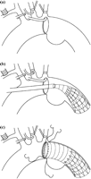
Procedure for conventional transaortic stent-grafting. A hemicircular aortotomy was performed on the anterior wall of the proximal arch aorta under selective cerebral perfusion and hypothermic systemic circulatory arrest (a). A sheath catheter was inserted into the descending thoracic aorta via a small aortotomy and advanced beyond the distal end of the aneurysm. The distal end of the stent-graft was adequately positioned under TEE guidance (b). The proximal end of the stent-graft was sutured onto the posterior aortic wall intraluminally and the anterior aortic wall transmurally through the aortotomy. The aortotomy was closed with the anterior wall of the proximal end of the stent-graft (c).
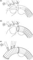
Procedure for hybrid transaortic stent-grafting. The aortic arch was transected proximal to the left subclavian artery and a stent-graft was inserted into the descending aorta (a). The proximal end of the stent-graft was sutured circumferentially to the transected aortic arch (b). The ascending and the proximal aortas were replaced with a branched graft, which was anastomosed to the proximal end of the stent-graft (c).
Every survivor underwent post-operative CT scans every 6 months. The excluded aneurysm space was evaluated by measuring the sizes of the outer and inner diameters of excluded aneurysms in CT slices, and compared to the same level CT slices of differing time periods (Fig. 3) . The maximum aneurysmal diameter was also measured at the same level CT slices. Shrinkage of the excluded aneurysmal space for each CT slice was serially calculated by comparison with a control CT slice taken a month after surgery (Fig. 3).
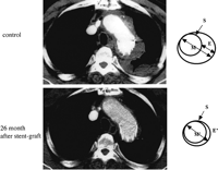
Shrinkage of the aneurysmal diameter as measured by transectional CT slices. The excluded aneurysmal space was evaulated by measuring the diameter of the aneurysm, not including the diameter of the stent-graft at 1 month (control) after surgery and each follow-up period. Percent shrinkage of the excluded aneurysm was calculated by comparing the diameter of the aneurysm at its maximum size (M, at 1 month; M′, at 26 months after stent-grafting) to the excluded aneurysmal space (E, at 1 month; E′, at 26 months after stent-grafting), which was the space excluded by stent-grafting (S, stent-graft) at each period.
Results were recorded as means±SD, and statistical significance was calculated using Student's t-test for paired observations. A P-value of less than 0.05 was considered to be statistically significant.
Stent-grafting was approved by the Hiroshima University Hospital Institutional Review Board for Human Studies, and performed after signed informed consent forms had been obtained from all the patients.
3 Results
Twenty-nine cases received stent-graft insertion via a small proximal aortic arch incision (conventional transaortic stent-graft); five stent-grafts were inserted through a circumferential distal aortic arch amputation and concomitant total arch replacement (hybrid transaortic stent-graft). The size of the stent-graft ranged from 24 to 34 mm in diameter. Selective cerebral perfusion and extracorporeal circulation times ranged from 46 to 86 min (mean 62 min) and 88 to 142 min (mean 116 min), respectively. Total operating time ranged from 225 to 402 min (mean 302 min). There were two hospital deaths due to a cerebral embolism during retrograde perfusion via the femoral artery and a peri-operative myocardial infarction (2/34; mortality 5.9%). One patient had received a conventional transaortic stent-graft, while the other had undergone a hybrid transaortic stent-graft. Thirty-two cases (94%) were discharged, but one case died from pneumonia a year later, and one hybrid stent-graft case suffered post-operative paraplegia. Paraplegia seemed to stem from a left vertebral arterial embolism, as there were numerous atheromatous plaques around the left subclavian arteric orifice (morbidity 2.9%). The remaining 31 cases survived without any serious complications during the follow-up period (Table 1) .
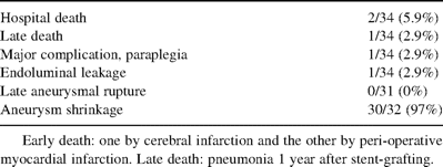
Results of the transaortic stent-grafting
Intraoperative transesosphageal echocardiography revealed endoluminal leakage in 1 of the 34 cases (2.9%). Stent-graft endoleakage persisted into the excluded aneurysmal space during the follow-up period (Table 1); an additional stent-graft was applied, but inappropriately deployed. Since the aneurysmal diameter was growing very slowly (2 mm/year) and the risk of aneurysmal rupture was thought to be low, this patient was observed without re-operation during the follow-up period.
Post-operative serial CT examinations were available in 31 cases and continued during the follow-up period. CT revealed extreme excluded aneurysmal space shrinkage in 30 of 31 cases, excluding the case with endoluminal leakage. Changes in maximum aneurysmal diameters decreased significantly from 6.7±1.8 cm (at discharge) to 5.4±1.3 cm (at 12 months) and 4.4±1.3 cm (at 24 months) (Fig. 4) . Excluded aneurysmal space size decreased significantly from 100% (at discharge) to 61% (at 12 months) and 36% (at 24 months) post-operatively (Fig. 5) . Amongst 30 patients with successful thromboexclusion, 9 patients (9/31, 29%) showed extreme excluded aneurysmal space shrinkage that disappeared or vanished during the follow-up period (Fig. 6) . Aneurysm sizes regressed in all the thrombo-excluded patients from the third month onwards of stent-grafting. The case with endoluminal leakage showed gradual aneurysmal dilation during the follow-up period. There were no aneurysmal ruptures in the 31 cases who survived during follow-up, which ranged from 10 to 72 months (average 38.3±27.2 months). Latent neurological disorders, such as cerebral embolism or peripheral embolism were not observed.
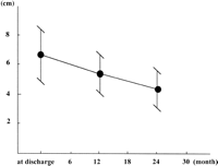
Changes in the maximum aneurysmal diameter. The maximum aneurysmal diameter decreased significantly from 6.7±1.8 cm (at discharge) to 5.4±1.3 cm (at 12 months) and 4.4±1.3 cm (at 24 months).
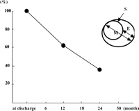
Percent shrinkage of the excluded aneurysmal space. Excluded aneurysmal spaces significantly decreased after successful stent-graft placement. We compare the size of the excluded aneurysmal space after 1 month (at discharge) with the size of the excluded aneurysmal space at each follow-up period. The percent shrinkage of the excluded aneurysmal space: the excluded aneurysmal space at each follow-up period/the excluded aneurysmal space at 1 month ×100 (%). M, maximum aneurysmal diameter; S, stent-graft; E, excluded aneurysmal space.
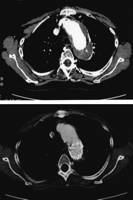
Complete disappearance of the excluded aneurysmal space. This patient showed extreme shrinkage of the excluded aneurysmal space, which vanished completely during the follow-up period (14 months after stent-graft).
4 Discussion
Parodi et al. initially reported successful placement of abdominal aortic aneurysmal endovascular stent-grafts [7]. When a transcatheter endovascular stent-graft is considered for a patient with a thoracic aortic aneurysm, many anatomical and technical problems must be addressed. In particular, anatomical suitability is important and usually makes endoluminal device deployment difficult because of the severely curved aortic arch configuration and the risk of cervical arterial branch obstruction [2,3]. Our small transcatheter stent-grafting experience also showed difficulty of complete distal aortic arch aneurysmal exclusion when the aneurysm extended to the aortic arch, easily causing endoluminal leakage in the site proximal to the stent-graft. In addition, many distal aortic arch aneurysm patients also required coronary artery bypass grafting or additional cardiac surgeries. Therefore, we performed transaortic endovascular stent-grafting for distal aortic arch or proximal descending thoracic aortic aneurysms, instead of prosthetic replacements via left thoracotomies [6]. We performed subclavian–subclavian arterial bypasses with small prostheses in five cases because during extracorporeal circulation, prostheses were used as arterial perfusion conduits. We think translocation of the left subclavian artery to the left carotid artery was better than prosthetic bypass.
We modified the original transaortic endovascular stent-grafting procedure and enhanced it by decreasing the aortic arch incision. We can introduce the stent-graft through a small aortic arch incision between the left common carotid artery and the left subclavian artery. This method has several advantages, such as a decrease in bleeding from the small aortotomy, a shorter operative time, and no recurrent nerve damage. In fact, this procedure was performed without blood transfusion. Moreover, the risk of distal stent-graft displacement is nullified by suturing the stent-graft's proximal. From our transcatheter stent-grafting experience in abdominal aortic aneurysms, some stent-grafts dislocate post-transcatheter placement.
There was one serious cerebral embolism and one peri-operative myocardial infarction. Both cases seemed to occur from aneurysmal embolism. Antegrade arterial perfusions were principally accomplished via ascending aorta or right subclavian arteries. However, transfemoral arterial perfusion was used in two patients because of calcified ascending aortas, which may cause an embolism by retrograde perfusion. Another case resulted in paraplegia because the aneurysm was severely atheroscleric; the left subclavian artery had atheroscleroric plaques, which seemed to have caused thromboembolism in the left vertebral artery. Post-operative mortalities and morbidities were the same as those in conventional prosthetic total arch replacements, meaning that this procedure is under progression, and requires further refinement. Peri-operative parameters, such as perfusion time and blood loss were lower than those in conventional prosthetic total arch replacements.
Follow-up CT examinations revealed that the 30 successful cases demonstrated significant excluded aneurysmal space and several cases showed complete aneurysmal disappearance. These CT findings confirmed complete aneurysmal exclusion by this procedure, without risk of aneurysmal rupture after successful transaortic [8,9] and transcatheter [10] stent-graft placements. We compared aneurysm diameters during follow-up with those measured a month after surgery, as we intended to evaluate excluded aneurysmal space shrinkages. In general, excluded aneurysmal space diameters tended to reduce, but not immediately; diameters dilated occasionally and began to shrink later on, from 1 to 6 months after transcatheter stent-grafting [1–3]. Our successful cases also showed excluded aneurysm shrinkage 2–3 months after surgery. Shrinkage seemed to be more rapid than in transcatheter endoluminal stent-grafting. We think this is a specific characteristic of transaortic stent-grafting because it results in complete endovascular coverage when the proximal stent-graft end is sutured.
4.1 Limitations
There are several limitations to this procedure. One is the requirement of a stent and a sheath catheter. Others include the occurrence of paraplegia and endoluminal stent-graft leakage. The mechanism for paraplegia after transaortic stent-grafting is unknown. However, we think it occurs by embolization of the left vertebral or critical intercostal arteries.
5 Conclusion
Although there are several caveats indicating the prevention of neurological complications, transaortic stent-grafting is a feasible treatment for true thoracic aortic aneurysms.
Dr J. Bachet (Paris, France): Just for my information. I noticed that in some patients you have done a right to left subclavian-subclavian bypass. Why do you prefer to do that instead of a simple translocation of the left subclavian artery into the left carotid artery? Don't you think that a transverse bypass is more complicated and more prone to thrombosis and could preclude going back through the sternum?
Dr Sueda: I agree with you. But unfortunately, I don't know the exact procedure to anastomose directly the left subclavian artery to the left carotid artery. So I will try it next time.
Dr M. Krason (Zabrze, Poland): I would like to ask you what is your approach for positioning of the stent-graft during open procedure? Do you use an X-ray or just you position it based on previously performed CT scan?
Dr Sueda: We only perform it through the median sternotomy. We measure the size of the stent-graft preoperatively by CT and during operation by transesophageal echocardiography as well as the site of placement was decided by transesophageal echocardiography.
