


Abstract
A case of thoraco-abdominal aortic aneurysm complicated after permanent clamping of the descending aorta (thromboexclusion) is reported. Angiographic and operative findings were: (1) a pseudo-aneurysm right at the distal anastomosis of previous intrathoracic bypass for pseudo-coarctation of the aorta filled by left ninth intercostal artery, which was supplied by the left internal thoracic artery; and (2) the cervical and thoracic spinal cord were supplied by the left vertebral artery and the mediastinal branch of the left thyrocervical trunk. This rare cause of a thoraco-abdominal aortic aneurysm and the significance of the subclavian artery as a source of spinal cord blood supply are discussed.
1 Patient
A 65-year-old woman was admitted with the diagnosis of a thoraco-abdominal aortic aneurysm. She had a history of multiple surgeries performed at a community hospital under the diagnosis of aortitis (Fig. 1a) . At the age of 31 years, stenosis of the descending aorta, pseudo-coarctation of the aorta, was bypassed from the distal aortic arch to the descending aorta. At the age of 51 years, graft occlusion of the intrathoracic bypass and a pseudo-aneurysm at the proximal anastomosis were treated by permanent clamping of the descending aorta and ascending aorta to the abdominal aorta bypass (flow reversal thromboexclusion method [1]). When she was 60 years old, an aneurysm of the thoraco-abdominal aorta was observed. The arch to the descending aorta bypass graft was closed and a bypass from the ascending to bilateral external iliac arteries was installed. The diameter of the aneurysm decreased from 63 to 42 mm in the next 2 years, however, it increased again to 58 mm in the following 3 years. She was referred to us for further examination and surgical treatment.
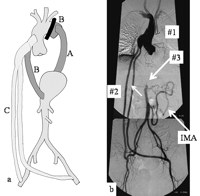
(a) History of multiple surgeries. (A) At the age of 31 years. (B) At the age of 51 years. (C) At the age of 60 years. (b) Preoperative aortography: (#1) total clamping of the descending aorta; (#2) patent bypass from the ascending aorta to bilateral iliac arteries; and (#3) occlusion of the abdominal aorta just above renal arteries. IMA, inferior mesenteric artery.
She was asymptomatic and blood pressure was under control with a β-blocker and a vasodilator. The ankle/ brachial pressure index was 1.0. Laboratory data were within normal limits including C-reactive protein at 0.31 mg/dl without the administration of steroids. The erythrocytes sedimentation rate was moderately accelerated, 25 mm at 1 h. Aortography revealed total occlusion of the descending aorta and a patent ascending aorta to bilateral iliac arteries bypass. Retrograde flow in the abdominal aorta supplied the inferior mesenteric artery and bilateral renal arteries. The abdominal aorta was occluded just above the renal arteries. The superior mesenteric artery and celiac trunk were not visualized. The collateral flow from the inferior mesenteric artery supplied the upper abdomen (Fig. 1b).
As the aortography did not demonstrate the source of blood flow into the thoraco-abdominal aortic aneurysm and spinal cord blood supply, selective angiography and MR angiography [2] were performed. The left eighth and ninth intercostal arteries were visualized from the left internal thoracic artery (LITA; Fig. 2a) . The ninth intercostal artery supplied fine collateral to thoraco-abdominal aortic aneurysm and left staining of the aneurysm (Fig. 2b). The upper portion (cervical to upper thoracic) of the anterior spinal artery was visualized from the left vertebral artery. The mediastinal branch of the thyrocervical trunk provided abundant collateral flow and supplied intercostal arteries. From the left sixth intercostal artery, the anterior spinal artery was visualized through a hairpin-curved radicular artery (Fig. 2c). On MR angiography, a hairpin-curved radicular artery branching out from the right 11th intercostal artery to the anterior spinal artery was diagnosed as Adamkiewicz artery.
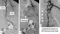
Selective angiography. (a) The left eighth (VIII) and ninth (IX) intercostal arteries were visualized from the LITA. (b) The ninth intercostal artery supplied fine collaterals to the thoraco-abdominal aortic aneurysm and left dye staining. (c) From the left sixth intercostal artery supplied by a mediastinal branch of the thyrocervical trunk, an anterior spinal artery (bold arrows) was visualized through a hairpin-curved radicular artery (thin arrows).
The chest was opened through the left seventh intercostal space and adhesions were carefully dissected. A cerebrospinal fluid drainage [3,4] was placed the day before the operation, and motor evoked potential (MEP) was monitored during the operation. The left eighth rib was incised together with the eighth intercostal artery. The distal portion of the LITA was clamped while confirming no change in MEP amplitude. When the aneurysm was incised, the bypass graft from the distal arch to the descending aorta was completely detached. The back flow from the left ninth intercostal artery was confirmed and closed inside the aneurysm. MEP showed no change during and after surgery.
The postoperative course was uneventful and the patient presented no symptoms of spinal cord injury. Twelve months after the operation, no recurrence of aneurysm formation was confirmed by CT.
2 Discussion
In the present case, the following issues were a matter of concern: (1) the source of blood flow into the thoraco-abdominal aneurysm; (2) spinal cord blood supply; and (3) approach to the aneurysm at surgery, which might require the ligation of left intercostal arteries and/or the internal thoracic artery which were the possible source of blood flow into the Adamkiewicz artery. The interventional coiling of intercostal arteries or the internal thoracic artery was not indicated as this possibility.
Edwards et al. [5] described that in patients with coarctation of the aorta, inflow into the collateral circulation is primarily from the branches of subclavian arteries. As in this patient blood supply from the aorta to upper and lower intercostal, upper lumbar, celiac trunk, and superior mesenteric arteries was absent, the branches of subclavian arteries were supposed to be a source of blood flow into the lower thoracic spinal cord.
Preoperative selective angiography of the branches of the left subclavian artery, MRA, and operative findings showed that the left vertebral artery supplied the cervical portion of anterior spinal artery; that the LITA did not supply Adamkiewicz artery; and that two major radicular arteries showing a hairpin-curved shapes were directly connected with the anterior spinal artery and were supplied by a highly developed mediastinal branch of the left thyrocervical trunk.
The right subclavian artery was not evaluated precisely, however, it could be emphasized that the left subclavian artery was a significant source of spinal cord blood supply.
