


Abstract
A case of benign sugar (clear) cell tumor of the lung with an unusual clinical presentation and its evaluation with computed tomography are reported. A 48-year-old man presented with one episode of hemoptysis. Chest radiographs revealed a round nodule in the lower left lung lobe, and fiberoptic bronchoscopy was normal. On the computed tomography scans, the nodule showed intense post-contrast enhancement (74.7 Hounsfield units). The patient underwent a left thoracotomy, and a segmentectomy was performed. Pathologic examination showed a benign sugar cell tumor of the lung. The patient is alive and has remained free of disease for the last 2 years. To the best of our knowledge, this is the first case report of sugar cell tumor located in lung parenchyma that presented with hemoptysis and the second report of the contrast-enhanced computed tomography findings in this neoplasm.
1 Introduction
The sugar (clear) cell tumors of the lung are rare benign neoplasms originally described in 1963 by Liebow and Castleman [1]. However, until this moment, only sporadic cases of this neoplasm have been reported in the world literature. The tumors are composed of a rich blood supply and clear cells with large amounts of intracytoplasmic glycogen, thus the denominations clear cell tumor and sugar cell tumor. The tumor cells show immunoreactivity for HMB-45 and S-100 and no reactivity for Cytokeratin 7, which establishes the definitive diagnosis [2]. This paper describes a benign sugar cell tumor of the lung (BSCT) with an unusual clinical presentation and the contrast-enhanced computed tomography (CT) findings in this neoplasm.
2 Case report
A 48-year-old white man presented to the emergency department with acute hemoptysis of 70 ml. On clinical interview, he had no previous diseases, but was a 30 pack-year smoker. Physical examination showed no abnormalities, and laboratory studies were unremarkable.
The chest radiographs showed a lower left lung lobe nodule. However, bronchoscopy did not reveal any abnormalities. Contrast-enhanced CT scan disclosed a 2 cm round nodule with a post-contrast enhancement of 74.7 Hounsfield units (43.7–118.4 Hounsfield units) (Fig. 1) .
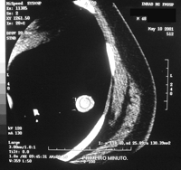
Contrast-enhanced computed tomography scan showing a 2 cm round nodule in the lower left lung lobe measuring 118.4 Hounsfield units.
The patient underwent a left thoracotomy, and a segmentectomy was performed. Grossly, the tumor was 2 cm in diameter, dark red and demarcated from the surrounding pulmonary parenchyma. The frozen section biopsy showed clear cells arranged in nests and in long cords. The nuclei were oval, and no mitotic figures were seen. The findings were consistent with BSCT, and the lobectomy was not performed.
The hematoxilin–eosin stain confirmed the above findings (Fig. 2) . The clear cytoplasm contained numerous glycogen granules as demonstrated by periodic acid-Schiff staining. The tumor was immunoreactive for S-100 and HMB-45 and nonreactive for carcinoembryonic antigen, cytokeratin 7 and 20, chromogranin and synaptophysin, characterizing the BSCT [2].
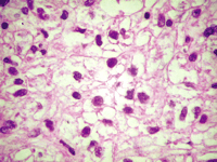
Microscopic findings. This section of the tumor shows cells with clear cytoplasm and regular nuclei in size. No mitotic figures were seen (H&E stain, original magnification 400×).
Further investigation showed no evidence of clear renal-cell carcinoma. The patient has been followed for 24 months since his surgery and is well, without evidence of recurrent or metastatic disease.
3 Discussion
There are <40 cases of BSCT reported to date in the English literature. Patients usually ranged in age from 40 to 60 years with equal sex prevalence. The youngest patient was an 8-year-old child. The typical clinical manifestation was incidentalomas on routine chest radiographs or CT scans. However, only one report described a patient with BSCT that presented with hemoptysis. The tumor was located in the upper trachea [3]. The biological behavior of this neoplasm was traditionally considered benign, although a 1976 report had described the case of a patient with BSCT presenting with blood-streaked sputum, who died from hepatic and peritoneal metastases 17 years after diagnosis [4]. The original features of the case presented here are hemoptysis as the first sign of a BSCT located in the lung parenchyma and contrast-enhanced CT findings.
Radiographically, the BSCT presents as rounded, smoothly countered, peripheral parenchymal nodules without evidence of cavitation or calcification. There are no specific lobar distributions. The main CT feature of this case is the intense post-contrast enhancement, consistent with a previous report [5]. This CT finding appears to be related to its rich vascular stroma as described in rounded atelectasis in workers exposed to asbestos [6].
The BSCT are usually diagnosed by thoracotomy. Only one report described the case of a patient with BSCT diagnosed pre-operatively by a transbronchial lung biopsy [7]. The patient underwent a thoracoscopic surgery without prior observation. Although observation may be an accepted method of management for benign pulmonary tumors, surgery is a good option of treatment for BSCT. Reports described a case of a patient who died from metastatic BSCT and another case of BSCT that doubled its diameter within 21 months [7]. The benign behavior is not invariable, and the aspects that indicate this behavior still need to be clarified. Our patient was a heavy smoker older than 40 years with a lung nodule with intense post-contrast enhancement as seen on CT scan, and was considered at high risk for a lung neoplasm [8]. He was submitted to lung segmentectomy after frozen section biopsy showed a BSCT. Other authors have successfully treated these benign tumors with sublobar resections, such as segmentectomy and wedge resection [7].
Grossly, the BSCT predominantly appears as well-circumscribed, peripheral, red-tan nodules measuring 3 cm or less in diameter. Cut surfaces are typically homogeneous and glistering without evidence of hemorrhage, necrosis, cavitation or calcification. One BSCT that metastasized presented as a lobulated, dark red, centrally necrotic mass measuring 4.5 cm in diameter [4].
The frozen section biopsy shows clear cells with oval nuclei, and no mitotic figures are seen. This technique may diagnose BSCT intraoperatively, avoiding unnecessarily extensive lung resections. The hematoxilin–eosin stain confirms the above findings, and the periodic acid-Schiff staining shows glycogen granules in cytoplasm of these clear cells, thus the name sugar cell tumor. The immunohistochemical-staining pattern of BSCT is unique, positive for HMB-45 and S-100 and negative for cytokeratin 7. The above findings indicate that BSCT exhibits melanocytic differentiation, distinguishing it from other clear cell neoplasms [2].
BSCT may be a very rare benign cause of hemoptysis [9,10]. Due to its intense post-contrast enhancement on CT scans, this tumor may simulate a malignant neoplasm, such as primary or metastatic lung cancer. Histologic and immunohistochemical findings differentiate this tumor from the metastatic renal cell carcinoma.
