-
PDF
- Split View
-
Views
-
Cite
Cite
A.D.L. Sihoe, C.S.K. Cheung, R.H.L. Wong, A.P.C. Yim, Giant pulmonary arteriovenous malformation, European Journal of Cardio-Thoracic Surgery, Volume 25, Issue 4, April 2004, Pages 648–649, https://doi.org/10.1016/j.ejcts.2004.01.002
Close - Share Icon Share
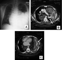
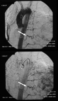
A 44-year-old man was admitted with coughing and dyspnea for 3 weeks. Chest radiography and thoracic CT scanning confirmed a huge left lung arteriovenous malformation (Fig. 1) . Due to a prohibitively high operative risk, he was investigated with pulmonary angiography in preparation for embolization therapy (Fig. 2) . The patient subsequently refused all further therapy.

Chest radiograph (A) and thoracic CT with intravenous contrast enhancement at the level of the pulmonary trunk bifurcation (B) and the lower thorax (C). There is a huge isodense soft tissue mass with speckles of calcification in the left lung. Enhancing serpiginous vessels characteristic of a pulmonary arteriovenous malformation are seen within the mass (arrowed). Prominent feeding vessels arising from the left main pulmonary artery favor a diagnosis of arteriovenous malformation rather than pulmonary sequestration, the major differential diagnosis. A component of the lesion with mixed fat and soft tissue content extends over the posterior mediastinum to the right hemithorax. The whole lesion causes gross displacement of the heart anteriorly and significant contralateral mediastinal shift.

Arch aortogram with pulmonary angiography illustrating the large pulmonary arteriovenous malformation. In addition to the pulmonary arterial supply, there are also feeding vessels from the left internal mammary, left bronchial, left phrenic, and at least four intercostal arteries, which are normally more typical of pulmonary sequestration. The feeding vessels are dilated and tortuous distally when approaching the lesion (arrowed).


