


Abstract
Objectives: Non-small cell lung cancer (NSCLC) tissue produces numerous growth factors, which are multifunctional and considered predictive of patient survival. This study sought to evaluate the relationship between concentrations of basic fibroblast growth factor (bFGF), vascular endothelial growth factor (VEGF), hepatocyte growth factor (HGF) in NSCLC tissue and clinicopathological factors, and determine whether these factors correlate with long-term survival. Methods: We retrospectively investigated 71 patients with histologically confirmed adenocarcinoma or squamous cell carcinoma of the lung, for whom the primary curative approach was surgery, and who received no chemotherapy or radiotherapy prior to surgery. bFGF, VEGF, HGF were measured in extracts prepared from these 71 frozen tissue samples by ELISA. Five- and 10-year survival was obtained to determine the most important predictors of long-term survival. Results: Among clinicopathological parameters, the mean concentration of bFGF was significantly higher in tissue extracts from cases involving metastatic nodal involvement (87.5±69.3 ng/100 mg protein) than in those without nodal involvement (57.6±42.5 ng; P<0.05). Levels of VEGF in adenocarcinoma (26.8±34.0 ng) were higher than for squamous cell carcinoma (12.2±13.8 ng; P<0.05). HGF levels also demonstrated histological differences (14.7±7.7 vs. 10.6±9.7 ng, P<0.05). bFGF protein levels were basically the same, but showed no statistically significant differences with respect to histological type (72.5±55.2 vs. 63.6±51.5 ng). Patients with high levels of bFGF or VEGF showed significantly worse survival rates than patients with low levels (P=0.0059; P=0.0134). In particular, high concentrations of both bFGF and VEGF correlated with markedly poor prognosis (P<0.0001). Multivariate analysis indicated that lymph node involvement and levels of bFGF and VEGF were independent prognostic factors in cases of NSCLC (adenocarcinoma or squamous cell carcinoma) involving patients who had undergone curative resection. Conclusions: Adenocarcinoma associated with lung cancer is regarded to have biological characteristics that distinguish it from squamous cell carcinoma. Node invasion may be associated with bFGF. bFGF and VEGF augment each other and are associated with leading to poor prognosis. The results of this study suggests that effective therapy to block angiogenesis may need to address at least both of these factors in cases of NSCLC.
1 Introduction
The prognosis for non-small cell lung cancer (NSCLC) has generally been poor, even with complete resection and post-operative adjuvant therapy. Angiogenesis regulates the growth and metastasis of solid tumors and has recently emerged as a therapeutic target for the development of anti-angiogenic agents for cancer treatment [1,2]. NSCLC tissue produces many growth factors, including epidermal growth factor (EGF), transforming growth factor-a, transforming growth factor-b, and platelet-derived growth factor (PDGF) [3,4]. These growth factors are involved in a wide range of activities, including promotion of mitogenesis, angiogenesis, chemotaxis, cellular differentiation, and tissue repair. Basic fibroblast growth factor (bFGF) is a secreted multifunctional cytokine and potent stimulator of angiogenesis [5,6]. Vascular endothelial growth factor (VEGF) also plays an important role in inducing endothelial cell proliferation and neovascularization [7,8]. Current reports indicate that serum VEGF in patients with lung cancer may serve as a useful indicator of tumor angiogenesis [9]. Hepatocyte growth factor (HGF), which is frequently over-expressed in NSCLC, plays multifunctional regulatory roles in growth. It also plays a significant role in tumor angiogenesis and metastasis [10,11]. However, their potential role in tumor progression remains unclear.
Many reports have evaluated the expression levels of these factors immunohistochemically. This review analyzes the results of clinicopathological studies on the prognostic role of angiogenic factor expression in NSCLC. But the criteria for cut-off points and stain methods differ from study to study. Standardizing angiogenesis assessment by microvessel count is difficult. To our knowledge, few investigators have reported on the relationship between actual tissue levels of angiogenetic factors and prognosis in NSCLC cases. In this study, we investigated levels of angiogenetic activity, bFGF, VEGF, and HGF in tissue extracts from cases of human lung cancer tissue. We attempted to clarify how these angiogenetic factors affect prognoses in cases of operable NSCLC, compared to earlier conventional methods.
2 Material and methods
2.1 Patients and methods
Specimens from 124 patients with primary NSCLC were obtained at surgical resection. Patients were excluded if they had undergone previous neoadjuvant chemotherapy, non-curative resection, or exhibited small cell lung cancer, adenosquamous, or other rare histology result. Patients who died within 1 month were also excluded. The study evaluated a final screened group of 71 patients who underwent curative operation (lobectomy or pneumonectomy with lymph node dissection) at the Second Department of Surgery of Fukuoka University Hospital, during the 7-year period from 1980 to 1988. Patient age ranged from 35 to 84 years (average age 67.1). The subject group consisted of 17 female and 54 male patients. Hematoxylin- and eosin-stained sections were subjected to histological diagnosis, grading, and nodal status assessment. Twenty-one patients had squamous cell carcinomas, while 50 patients had adenocarcinomas. Lymph node involvement was present in 29 cases. The breakdown by tumor sizes is: T1, 23 cases; T2, 44 cases; T3, 4 cases. Histological grade I was confirmed in 42 cases, II in 12 cases, and III in 17 cases. Table 1 shows the clinicopathological factors for 71 patients.
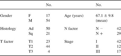
The clinicopathological factors of the 71 patients with NSCLC
The routine follow-up of these patients after surgery consisted of clinical evaluations performed every subsequent month. The resected NSCLC tissues were immediately stored at −80 °C until enzyme extraction. Sections of tumor remnants were stained with hematoxylin and eosin and subjected to histological examination. The clinicopathological parameters studied for the prognostic tissue levels of bFGF, VEGF, HGF were concentration, age, tumor size, lymph node involvement, histological type, and histological grade. All tumors were staged post-operatively, according to the classification system of the Internal Union Against Cancer (UICC).
2.2 Assay for bFGF,VEGF, HGF
Frozen tumor tissue (0.2 g) from each subject was quickly thawed at room temperature, minced, and rinsed with ice-cold 3 mM Tris–HCl buffer (pH 7.5) containing 250 mM sucrose. The minced portions were suspended in 1 ml of an ice-cold solution containing 3 mM Tris–HCl buffer (pH 7.5) and 250 mM sucrose, followed by homogenization in an ice bath with a glass homogenizer (ANEX 30420, Teraoka Co Ltd, Osaka, Japan). The homogenate was further homogenized with an ultrasonic homogenizer (USH-20Z20S, Tokyo Rikakikai Co Ltd, Tokyo, Japan) for 2 min at 5 °C and centrifuged at 3000 rev./min for 20 min at 5 °C. The supernatant was assayed for each enzyme concentration. The tissue extracts were stored at −80 °C until the assays. The bFGF concentration was measured using an enzyme-linked immunoabsorbent assay kit (R and I Systems Inc, USA), a three-step sandwich method. The detection limit of this assay is 0.1 ng/ml. To account for the possible influence of the extraction buffer, we compared standard curves with and without the extraction buffer. The protein contents of tissue extract were measured by the bicinchoninic acid method. All bFGF protein values are given in units of ng/100 mg protein. The VEGF concentration was also measured by an ELISA kit (Amersham Bioscience, USA). HGF was measured using an ELISA kit (Institute of Immunology Co Ltd, Tokyo Japan).
2.3 Statistics
Data are expressed as mean±SD. All statistical analyses were performed with the StatView software package (StatView 5.0.1, SAS Institute Inc.). Survival rates were calculated by the Kaplan–Meier method, while survival curves were compared using a log-rank test. For multivariate analysis, a Cox's proportional hazards regression model was used to evaluate variables that were significant predictors of survival. The threshold of significance was set at P<0.05. The cut-off values (high and low) were set at the median distribution of each factors.
3 Results
Table 2 shows extract concentrations of the 71 NSCLC for the 71 subjects.
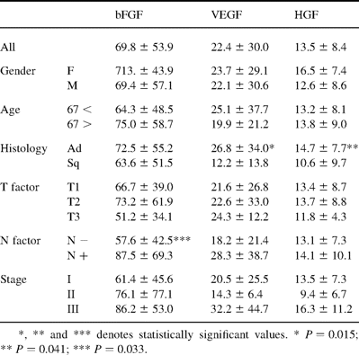
Relationships between concentrations of bFGF, VEGF, HGF and clinicopathological factors in 71 patients with NSCLC
3.1 Variation of bFGF
The bFGF concentration in NSCLC tissue was 69.8±53.9 ng/100 mg protein (range 4–174 ng/100 mg protein). No significant associations were observed between levels of bFGF and age, gender, T factor, or histological stage. There were no significant differences in histologic type, but adenocarcinoma was somewhat higher than in squamous cell carcinoma (P=0.26). Only node involvement was significantly associated with high levels of bFGF concentration (P=0.033).
3.2 Variation of VEGF
VEGF concentration in tissue was median 22.4±30.0 ng/100 mg protein (range 3–187 ng/100 mg protein). There were no significant associations between VEGF levels and age, gender, T factor, or histological stage. The only significant difference involved histologic type (P=0.015). But significant association with node involvement was not observed (P=0.174).
3.3 Variation of HGF
The median value for HGF concentrations in tissue extracts was 13.5±8.4 ng/100 mg protein (range 2–38 ng/100 mg protein). No significant association was observed between HGF levels and age, gender, node involvement, T factor, or histological stage. The only significant difference was in histologic type (P=0.041). Cases of adenocarcinoma showed higher levels than cases of squamous cell carcinoma. This histologic difference was basically the same as for VEGF and bFGF.
3.4 Relationship between extract levels of each factors and prognosis
A cut-off point was determined in our study to evaluate prognostic factors. This cut-off point for lower and higher quartiles of the total distribution of each factor activity was identified by the method of Tandon et al. [12]. The cut-off levels were 48 ng/100 mg protein in bFGF, 18 ng/100 mg protein in VEGF, and 13 ng/100 mg protein in HGF. Fig. 1a shows Kaplan–Meier survival plots generated from curves stratified by bFGF status. The 5-year survival of patients (n=38) with high bFGF was 29.0%; that of patients (n=33) with low bFGF was 70.4%. The 10-year survival of patients with high bFGF was 15.2%; that of patients with low bFGF was 49.4%. These were significantly associated with survival (P=0.0059). VEGF levels are shown in Fig. 1b. The 5-year survival of patients (n=28) with high VEGF was 36.7 vs. 66.7% of patients (n=43) with low VEGF. The 10-year survival of patients with high VEGF was 14.7 vs. 44.6% of patients with low levels. Statistically significant associations were found between survival and VEGF (P=0.0134). Fig. 1c also shows the basis for determining HGF levels. The 5-year survival rate for patients (n=31) with high HGF levels was 47.6%; the same rate for patients (n=40) with low HGF levels was 60%. The 10-year survival rate for patients with high VEGF levels was 20.8%; the same rate for patients with low VEGF levels was 41.3%. No significant correlation between HGF and survival was found in these cases of NSCLC.
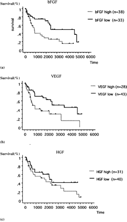
(a) Kaplan–Meier survival curves stratified for bFGF grade (high vs. low). H, high concentration; L, low concentration. Significant differences in outcome were observed between the high and low groups (P=0.0059). P-value was determined by the log-rank test. (b) Kaplan–Meier survival curves stratified for VEGF grade (high vs. low). H, high concentration; L, low concentration. Significant differences in outcome were observed between the high and low groups (P=0.0134). (c) Kaplan–Meier survival curves stratified for HGF grade (high vs. low). H, high concentration; L, low concentration. No significant difference in outcome was observed between high and low groups (P=0.2096).
We also analyzed the survival of the patients with each correlation factor. As shown in Fig. 2 , the 5-year survival rate for patients (n=15) with both high bFGF and VEGF levels was 13.0%. For all other combinations (high bFGF and low VEGF, low bFGF and high VEGF, and low bFGF and low VEGF), this rate was 64.9%. The 10-year survival rates for the respective groups were 6.5 and 42.9%. Statistically significant differences were observed between both high group and other group (P<0.0001). One other combination involving HGF factor levels alone did not correlate with the others. Fig. 3 shows the node status and survival curves. The 5-year survival rate for patients with node involvement was 32.1%; the rate for cases without node involvement was 62.8%. The 10-year survival rates for the respective groups were 12.9 and 42.8%. Statically significant differences were also seen between cases with and without lymph node invasion (P=0.0019).
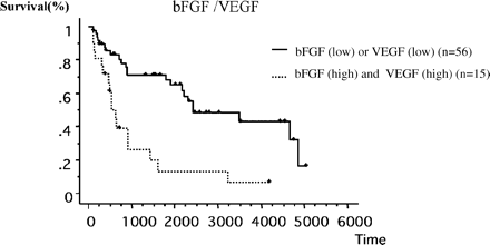
Kaplan–Meier survival curves stratified for co-high grade of bFGF and VEGF (high vs. low). H, co-high concentration; L, each low concentration. Co-high concentration group showed significant differences associated with poor outcome (P<0.0001).
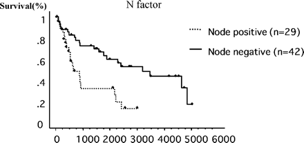
Kaplan–Meier survival curves stratified for node involvement (node negative vs. node positive). Significant differences were observed, which were associated with poor outcome (P=0.0019).
3.5 Prognostic significance of bFGF, VEGF, HGF tissue levels in cases of NSCLC, as determined by multivariate analysis
Multivariate analysis was performed to evaluate the respective prognostic roles of these tissue factors. All variables that significantly affected survival in analysis were used to draw up Cox proportional hazard models (Table 3) . After stepwise multivariate analysis, bFGF (P=0.0242) and VEGF (P=0.0428) maintained their independent prognostic effects on overall survival. Node status was another independent factor with significant effects (P=0.0205).
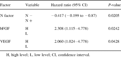
Significant variables in the log-rank test were computed from the multivariate Cox model analysis
4 Discussion
Many growth factors have been shown to be significantly related to non-small cell carcinoma and present in NSCLC tissue [3,4]. Most studies published in the literature agree that expressions of bFGF, VEGF and thymidine phosphorylase are important prognostic factors in cases of NSCLC [13]. Of these, bFGF is one of the most potent angiogenic factors. Recently, Joensuu et al. reported that high serum bFGF levels upon diagnosis are associated with poor outcomes for cases of lung cancer [14]. Our study showed no statistically significant differences in histology, gender, age, pathological stage with respect to bFGF obtained from tissue extracts. Lymph node progression, however, correlated with bFGF. We also recognized statistically significant differences in bFGF levels in relation to prognosis (P=0.0059). Another multifunctional protein, VEGF, showed no association with age, gender, or T factor. In contrast, we observed significant differences in histology (adenocarcinoma vs. squamous cell carcinoma). There were more node positive cases than node negative. Recently, VEGF subtype (VEGF-C or -D) has recently been shown to play a role which promotes the lymph node metastasis in colorectal cancer [15].
In contrast, some papers have reported that elevated serum bFGF in NSCLC cases is associated with good prognosis [16]. Another study reports that bFGF expression is associated with the absence of lymph node metastasis and less advanced cancer stages [17]. Mattern et al. found that bFGF association with angiogenesis was significant only in cases of VEGF co-stimulation [18]. Our data indicates that either autocrine or paracrine functions of bFGF and VEGF contribute significantly to lymphatic invasion, which is associated with poor prognoses. These data may suggest that both target therapies have a possibility of low chance of developing the recurrence after surgery.
We also investigated the relationship between HGF and other factors, but our findings did not suggest implications for prognosis, while levels of bFGF or VEGF were found to be related to poor outcomes in tissue from NSCLC cases. Research published by Fontanini et al. on a large-scale study of NSCLC cases indicates that increased nodal involvement is associated with microvessel count (MVD) [19]. Other studies have provided significant evidence that high MVD is one of the most important factors associated with NSCLC, inducing poor outcome after surgery [20,21]. However, each of these studies employ different cut-off points and staining methods (anti-factor VIII or CD31). MVDs are generally regarded as difficult to standardize, due to heterogeneous locations within tumors. New vessels are observed primarily in the tumor periphery. Several studies have demonstrated that bFGF or VEGF over-expression correlates with MVD and poor prognosis in NSCLC cases. In our study, methods of tissue extracts do not affect the tumor area. Histopathologies of adenocarcinoma tissue exhibit higher levels of these three factors than squamous cell carcinoma tissue. Differences were particularly marked for VEGF levels (P=0.015). And the HGF was also of the same difference between the histologies (P=0.041). These changes may be explained by the fact that squamous cell is locally extended, while adenocarcinoma is easily metastasized via microvessels. This data suggests difference in characteristics between squamous cell and adenocarcinoma in NSCLC cases. There are few reports of angiogenetic factors in the corresponding tumor homogenate from lung cancer cases. White et al. report that high levels of migration inhibitory factor (MIF) or VEGF from tumor homogenate are associated with increased risk of recurrence after resections involving lung cancer patients [22]. This is consistent with one of the outcomes observed in our research. HGF and its receptor Met are frequently over-expressed in non-small cell lung carcinomas, but their potential role in tumor progression remains unclear. Although we began our study by seeking to clarify HGF's role in poor prognoses in NSCLC, our unanticipated conclusion was that HGF was not associated with prognosis. Fuhrmann et al. report that plasma levels of HGF have been correlated with high plasma VEGF and metastatic disease [23]. But no relation was seen in our study between HGF and VEGF or bFGF. The angiogenic process is a complex multistep cascade under the control of positive and negative soluble factors. Each step represents a potential target for anti-angiogenic therapy. NSCLC are well known to be heterogeneous, with varying tumor cellularity and varying amounts of stroma. Tumor extracts' study informed us of each product instead of heterogeneous microenvironmental conditions in NSCLC. Takayama has demonstrated that adenovirus-mediated expression of a soluble VEGF receptor may inhibit tumor angiogenesis and suppress human lung cancer cell lines [24]. Elevated intracellular levels of bFGF are associated with resistance to chemotherapy in chronic lymphocytic leukemia (CLL) [25]. Multivariate analysis has confirmed bFGF and VEGF as well as node status to be an independent prognostic factor. We found that survival was significantly worse in cases demonstrating both bFGF and VEGF in tumor tissue than in cases evincing only one or neither factor. This data suggests that anti-angiogenic therapy must target not merely one, but both factors in patients suffering from NSCLC. Targeting both factors, as well as combined chemotherapy/radiotherapy, may result in more effective treatment.
