


Abstract
Malignant degeneration of neurogenic tumors has been reported to occur in 1–25% of patients with neurofibromatosis-I, and is the leading cause of cancer-related death in these patients. We report a case of multidisciplinary management of a giant malignant endothoracic nerve sheath tumor leading to histologically proven remission.
1 Introduction
Endo-thoracic tumors of neural origin are relatively rare [1]. Most of them are benign [1]. Of special interest are those tumors arising in patients suffering from neurofibromatosis (Von Recklingausen's disease). Neurofibromatosis I (NF-I) is the most common of the phakomatoses, or neurocutaneous disorders with an incidence of 1:2000–1:3000 [2]. It is due to an autosomal dominant disorder, involving chromosome 17, but it may be also consequent to a spontaneous mutation [3]. The thoracic manifestations of NF-I may be skeletal, pulmonary, and neurogenic [2]. Mostly, the neurogenic presentation of NF-I consists of focal or diffuse benign nerve sheath tumors [4]. However, from 1 to 25% of the tumors may have a malignant degeneration [3], and this is now termed malignant nerve sheath tumor [5].
We report a case of multidisciplinary management of a giant malignant endothoracic nerve sheath tumor.
2 Case report
A young student of 18 presented to the casualty of a district General hospital with generalized malaise, left-sided chest pain, and production of purulent sputum. He was a known case of NF-I diagnosed in his childhood. As the chest X-ray showed an ill-defined shadow involving the lower half of left chest, a diagnosis of left lower lobe pneumonia with para-pneumonic effusion was made (Fig. 1A) . A chest drain was inserted at this stage, but the drainage was very minimal. Intravenous antibiotics were started, temperature subsided and production of purulent sputum stopped. However, malaise and discomfort on the left side of chest persisted despite several course of IV antibiotics. The persistence of these symptoms led to further investigations, which also included a contrast CT-scan. This showed a large mass of heterogeneous attenuation involving the whole of left chest (Fig. 1B). A fine needle biopsy was at this stage performed suggesting a suspicion of neurofibrosarcoma. The case was at this stage discussed at the weekly multidisciplinary team meeting (MDT), attended by the senior respiratory physician, radiologist, oncologist, and thoracic surgeon. The planned strategy consisted of performing first surgery to be followed by adjuvant therapy. The patient was taken to the theatre and a left posterolateral thoracotomy was performed. A large, non-encapsulated tumor was found infiltrating the lateral portion of the diaphragm, the infero-lateral chest wall with protrusion through the 7th intercostal space. The entire lower lobe was incorporated in the tumor and the mass was adherent but not infiltrating the upper lobe. As expected tumor free resection margin was not achievable particularly at the diaphragm and laterally on the chest wall. An extensive debulking procedure was carried out. First, an extra-pleural approach was performed extended to the mid and lower left chest. A left lower lobectomy was then performed. No formal resection of the diaphragm or the chest wall was carried out, although the macroscopically evident tumor was extensively excised.
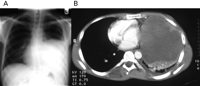
(A) Baseline plain chest X-ray; and (B) baseline contrast CT-scan.
The histopathology from the main mass showed high grade malignant peripheral nerve sheath tumor (neurofibrosarcoma) with the presence of densely cellular spindle cell sarcoma with high mitotic figure and extensive necrosis. Metastatic neurofibrosarcoma was also found in local lymph nodes. Multiple biopsies from the infiltrated parts of the diaphragm and chest wall showed infiltration with malignant cells. The tumor was staged (AJCC Staging) to G3 T3 N1 M0.
Post-operative recovery was free of major complications and the patient was referred to the oncologist as previously planned. Over the following 6 months, the patient underwent a total of six cycles of combination chemotherapy consisting of intravenous infusion of 42 mg of Doxorubicin and 1.67 mg of Ifosfamide every 3 weeks. During this period, CT scans were repeated every 2 months, showing no evidence of recurrence and, in fact, a gradual resolution of the thickening at the left costo-diaphragmatic angle. On completion of the chemotherapy, with a CT scan showing no more than residual pleural thickening in the lateral and posterior aspect of the left hemithorax, the patient underwent consolidation radiotherapy to the left hemithorax. (50 Gy of 6 MV photon iradiation in 25 fractions over 35 days).
At this stage, the case was discussed again at the weekly MDT meeting. The planned strategy consisted of a ‘second look thoracotomy’ with both diagnostic and curative intent. The patient and his parents (of whom one happened to be a physician) also preferred this option. On admission, chest X-ray and MR scan were unremarkable (Figs. 2A,B , respectively). At re-entry, no suspicious mass was identified, and multiple biopsies were obtained from lung, pleura, diaphragm, and skeletal muscle, particularly at the site of previous infiltration. Histology revealed no evidence of persisting malignant peripheral nerve sheath tumor. The patient was discharged on post-operative day 6. Further CT scans at 6 and 12 months follow up showed no evidence of recurrences.
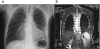
Plain chest X-ray (A); and MRI scan (B) following six cycles of chemotherapy and consolidation radiotherapy.
3 Discussion
Patients afflicted by NF-I are at increased risk of developing nervous system neoplasms including plexiformneurofibroma, optic glioma, ependymoma, meningioma, astrocytoma, and pheochomocytoma [2].
Mostly, the thoracic neurogenic presentation of NF-I consists of focal or diffuse benign nerve sheath tumors [4]. However, from 1 to 25% of these tumors may have a malignant degeneration [2]. As most benign neuronal tumors in patients with NF-I are asymptomatic, pain is a worrisome finding in this population, often indicating malignant degeneration [2]. Malignant change in preexisting superficial neurofibroma is easily detactable by rapid increase in size. But malignant change in a deep-seated lesion is a diagnostic challenge, and often pain may be the first sign, as happened to be in our patient.
There is very little in the literature with regard the treatment and the mid-term prognosis of such condition. Bruckner et al. [6] reported the case of a patient with neurofibromatosis developing a large inoperable malignant schwannoma on the posterior neck. The tumor underwent complete local regression following combined-modality treatment with radiotherapy, vinblastine, and doxorubicin. In keeping with this result, our case confirms the important role of chemo-radiotherapy in determining early complete local regression. In our case, however, the chemo-radiotherapy followed an extensive surgical debulking of the tumor, and the remission post chemoradiotherapy was confirmed with open multiple biopsies. With this strategy the patient remain under remission at 1 year as suggested by late imaging. The long-term prognosis of such treatment however remains unknown.
It is difficult to quantify the contribution of the MDT meeting to the outcome of this case. The first MDT consisted of a prolonged debate focusing on the patient’ age, the extension of the disease on the CT-scan and the very little literature available. Taking account of all these factors, it was decided to plan surgery first followed by adjuvant therapy. The idea was of removing most of the tumor first while achieving complete histology and staging as this might have facilitated the following chemo-radiotherapy. The extensive accurate debulking procedure might have been, indeed, of key importance in determining the success of the following chemotherapy and radiotherapy treatment.
Another critical decision in the management of this case was taken still in the context of a MDT meeting with regard the ‘second look thoracotomy’. Particularly, two options were debated at this time. The first consisted of a ‘wait and watch’ strategy inclusive of serial CT/MRI with a view of new cycles of chemo-radiotherapy in case of recurrence. The second option was more aggressive and consisted of a ‘second look thoracotomy’ with both diagnostic and curative intent with a view to removing any residual tumor if present and to perform multiple biopsies. It was also decided that in case of evidence of residual tumor a further cycle of chemo-radiotherapy was to be planned. The rationale in deciding to perform a second look thoracotomy rather than a ‘wait and watch’ strategy was that despite the very encouraging late results at MRI (Fig. 2), compelling evidence was needed prior making any decision in stopping the chemotherapy and radiotherapy treatments. To this end, multiple biopsies were taken during surgery under direct vision particularly at the left costo-diaphragmatic angle, the site showing infiltration of the tumor at first thoracotomy. It remains to be said that in making this decision we were facilitated by the patient age and by the very good cardio-respiratory status. Should these conditions have been poor, we would have obviously opted for a ‘wait and watch’ strategy.
It remains to be said that the absence of tumor at second operation is no safe guard against recurrence. To this end, we are performing every 6 months CT/MRI scans. Periodic CT scan or MRI has the advantage of diagnosing the recurrence at an early stage as development of heterogeneous attenuation or signal intensity [1,2,4]. This might increase the chances of success of a further cycle of chemo-radiotherapy if needed.
In conclusion, our case shows that an aggressive multidisciplinary management of giant malignant endo-thoracic nerve sheath tumor may lead to promising early result. More data are needed, however, to ascertain the mid- and long-term efficacy of such management.
