


Abstract
Objectives: Surgical radiofrequency ablation is increasingly used during open heart surgery for the treatment of chronic atrial fibrillation. The purpose of this study was to determine the effects of application of radiofrequency on coronary endothelial function and structure and establish the relationship between coronary lesions and distance of radiofrequency application. Methods: Six Landrace swine (25.9±2.0 kg) were included in the study. With the heart kept beating, three epicardial radiofrequency lesions (20 W, 20 s duration, 60 °C) 2 cm in length each, were created 1, 5 and 10 mm away from the left anterior descending and the right coronary arteries. The circumflex artery served as control. Coronary rings were placed in organ chambers. After contraction to KCl and prostaglandin F2α, endothelium-dependent relaxations to bradykinin were studied. Gomori trichrome and hematoxylin–eosin safran staining were used for histological evaluation. Results: Exposure to radiofrequency 1 mm from the coronary arteries caused a significant decrease in endothelium-independent contractions to KCl and endothelium-dependent relaxations to bradykinin compared to controls (P<0.05). No significant decrease of endothelium-dependent relaxations occurred for rings exposed to radiofrequency at a distance of 5 and 10 mm, compared to controls. Histological examination showed endothelial disruption and medial smooth muscle cells at different stages of necrosis up to 5 mm from the radiofrequency application site. Conclusions: Radiofrequency may induce coronary endothelial functional and morphological damages when applied less than 5 mm from the artery. Caution must be exerted during left atrial radiofrequency application due to the proximity of the circumflex artery.
1 Introduction
Chronic atrial fibrillation can be surgically treated by segmenting the atrial wall with linear lesions which were created in the early experience by complex mechanical incisions and sutures, according to the Cox-Maze procedure [1]. More recently, several studies have reported techniques by which transmural incisions have been successfully replaced by linear ablative lesions induced by the delivery of radiofrequency (RF) energy inside the left atrium [2–4]. This technique is also time-sparing compared to surgical incisions [5]. Pathological examination of the ablated myocardial zone usually show edema and necrosis of the myocardium, of variable extension and severity [6,7], and coronary arteries may be involved in the process. Previous experimental data demonstrated coronary artery obstruction adjacent to RF current lesions 48 h and even at a distance in time (>6 months) after RF current application [8]. However, to the best of our knowledge, no studies have looked at the effects of RF energy on the endothelial function of the coronary arteries, which controls the regulation of vascular tone and coagulation [9,10], with the attendant risk of spasm and thrombosis in case of major impairment.
During RF Maze procedures, some lesions are made at close proximity to the coronary arteries, especially the circumflex [11,12]. The purpose of this study was to determine the effects of application of RF energy induced by handheld probes on coronary endothelial function and structure, and establish the relationship between coronary lesions and the distance of RF application from the coronary artery.
2 Materials and methods
2.1 Radiofrequency probe and generator
Epicardial RF lesions were produced using an unipolar probe (Biosense Webster Inc., Johnson and Johnson, Diamond Bar, CA) coupled with a holder and a Ringer lactate continuous irrigation system (Fig. 1) . The electrode tip had a width of 2.3 mm. The radiofrequency current was delivered between the probe electrode and a large diathermy plaque positioned on the back of the animal. Each lesion was performed using a radiofrequency generator (Fig. 2) with a power output of 20 W (Dr. Osypka GmbH Medizintechnik, HAT 200S, Rheinfelden-Herten, Germany) during 20 s.
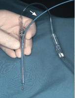
Probe (arrow) used in this experiment (Biosense Webster Inc., Johnson and Johnson, Diamond Bar, CA) coupled with a holder and a Ringer lactate continuous irrigation system. The electrode tip had a width of 2.3 mm.
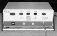
Radiofrequency generator (Dr. Osypka GmbH Medizintechnik, HAT 200S, Rheinfelden-Herten, Germany).
2.2 Experimental surgery
Six Landrace swine of either gender, aged 8±1 weeks and weighing 25.9±2.0 kg, were included in this study. Animals were maintained and tested in accordance with the recommendations of the Guidelines on the Care and Use of Laboratory Animals issued by the Canadian Council on Animals. The animals were sedated with an intramuscular injection of 25 mg/kg of ketamine hydrochloride (Ayerst veterinary Laboratories, Guelph, ON) and 10 mg/kg of xylazine (Boehringer Ingelheim, Burlington, ON), intubated and mechanically ventilated with an oxygen/air mixture (3:2). Anesthesia was maintained with 2.5% halothane inhalation (Halocarbon Laboratories, River Edge, NJ). The electrocardiogram was recorded from three subcutaneous limb electrodes. The heart was then exposed via a median sternotomy approach.
For each swine, three epicardial radiofrequency lesions (20 W, 20 s duration and 60 °C temperature), 2 cm in length were made with a slow mobilization of the tip of the probe on the right side of the left anterior descending artery (LAD) respectively at 1, 5, and 10 mm away from the artery (Fig. 3) . Three lesions were also created on the ventricular side of the right coronary artery (RCA). The left circumflex artery (LCX) served as control. The heart was then excised and placed in a cold modified Krebs-bicarbonate solution (composition in mmol/l: NaCl 118.3, KCl 4.7, MgSO4 1.2, KH2PO4 1.2, glucose 11.1, CaCl2 2.5, NaHCO3 25, and ethylenediaminetetraacetic acid 0.026).
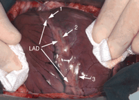
Three epicardial radiofrequency lesions (20 W, 20 s duration and 60 °C temperature), 2 cm in length made on the right side of the left anterior descending artery (LAD), respectively, at 1 mm (1), 5 mm (2), and 10 mm (3) away from the artery.
2.3 Organ chamber experiments
Coronary arteries were dissected free of the surrounding tissue in a Petri dish filled with cold modified Krebs-bicarbonate and were divided into rings 4 mm in length. Six rings were obtained from the RCA, and six from the LAD (two for each group). Two control rings were obtained from the LCX. All rings were placed in organ chambers (Emka Technologies Inc., Paris, France) filled with 20 ml of modified Krebs-bicarbonate solution heated at 37 °C and oxygenated with a carbogen mixture (95% O2 and 5% CO2). The rings were suspended between two metal stirrups with the upper one connected to an isometric force transducer. Data were collected with a biological signal data acquisition software (IOX 1.203; Emka Technologies).
Each arterial ring was stretched to the optimal point of its active length-tension curve as determined by measuring the contraction to potassium chloride (KCl) 30 mmol/l at different levels of stretch (stretch for optimal response was usually 3.5 g). The maximal contraction of rings was then obtained with addition of potassium chloride (KCl 60 mmol/l). After obtention of a plateau, all baths were washed twice with modified Krebs-bicarbonate solution and indomethacin (10−5 mol/l to exclude production of endogenous prostanoids), propranolol (10−7 mol/l to prevent the activation of β-adrenergic receptors) and ketanserin (10−6 mol/l to block serotonin 5-HT2 receptors) were added in each bath.
After 45 min of stabilization, prostaglandin F2α (PGF2α, range 2×10⁻⁶ to 3×10⁻⁵ mol/l) was added to obtain a contraction averaging about 50% of the maximal contraction to KCl. Endothelium-dependent relaxations to bradykinin (BK; an agonist which binds to receptor coupled to Gq-proteins) at incremental concentrations (10−12 to 10−6 mol/l) were recorded. At the end of the experiment, endothelium-independent relaxations were studied using a bolus of sodium nitroprusside (SNP 10−5 mol/l).
2.4 Histological analysis
Histological studies were done on animals killed 5 min after the application of the RF epicardial lesions. The heart was excised rapidly and placed in a formalin solution during 24 h. Paraffin blocks were molded from each lesion and three microscopic sections were made in the plane perpendicular to the long axis of the RF ablation zone for each lesion. Gomori's trichrome and hematoxylin–eosin safran (HES) staining were used, to evaluate the coronary vessels and surrounding myocardium.
2.5 Statistics
Continuous variables are expressed as mean±standard error of the mean (SEM). Preliminary two-way repeated measures analysis of variance (ANOVA) showed interaction between group and bradykinin concentrations. Due to this interaction, one-way repeated measures ANOVA was used to analyze the data. Contrasts were built to make predetermined comparisons of the means (Fig. 4) . One-way repeated measures ANOVA and contrasts was also used to analyze the data of Table 1 . Differences were considered statistically significant if the P-value was less than 0.05.

Cumulative concentration–relaxation response curves to bradykinin (BK) in porcine coronary arteries rings submitted to the radiofrequency lesions (RF) at different distances from the artery, and in controls rings. A P-value of less than 0.05 was considered statistically significant. PGF2a=PGF2α.
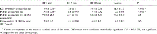
Amplitude of contraction to potassium chloride (KCl 60 mmol/l) and prostaglandin F2α (PGF2α) with the concentration used for all groups studied
3 Results
3.1 Coronary reactivity studies
3.1.1 Contractions
The amplitude of the contraction to KCl (60 mmol/l) and to PGF2α (range 2×10⁻⁶ to 3×10⁻⁵mol/l) was recorded for all groups (group 1, 5, 10 mm, and controls). There was a statistically significant lower amplitude of contractions to KCl and PGF2α in the rings exposed to RF at 1 mm, compared to the other three groups. There was no statistically significant difference in contractions to KCl and PGF2α between the rings exposed to RF at 5 and 10 mm, when compared to the control group (Table 1).
3.1.2 Relaxations
3.1.2.1 Endothelium-dependent relaxations
There was a statistically significant decrease (P<0.05) in endothelium-dependent relaxations to BK in rings exposed to RF at 1 mm, compared with the control group and the others groups. There was no statistically significant decrease of relaxations to BK between rings exposed to RF at 5 and 10 mm, compared with the control group (Fig. 4).
3.1.2.2 Endothelium-independent relaxations
No statistically significant differences in relaxation to SNP was observed between groups with all rings achieving 100% relaxation (data not shown).
3.2 Macroscopy and histology
Application of radiofrequency energy caused a pale yellow discoloration of the epicardium in an area extending 5 mm around the tip of the ablation probe, inducing a 10-mm wide rigid lesion 20 mm in length (Fig. 3).
After formalin fixation, the lesions were readily visible on cut sections. The lesions were stiff and pale compared to the surrounding myocardial tissue. Microscopic examination showed a well delimited RF lesion on the myocardium. The borders of these lesions were precisely identifiable with the Gomori Trichrome stain. The depth of lesions was 5±1 mm (Figs. 5 and 6) . At the center of the RF lesion, in a radius of 4 mm around the application point, myocardial cells showed complete coagulation necrosis surrounded by edema and hemorrhage. At the periphery of the lesion, contraction band necrosis was visible at a depth of 1 mm in depth encircling the previously described area of coagulation necrosis. The section of LAD artery located away from the zone of RF application, was morphologically normal (Fig. 7A) . The section located inside the RF effect area (Fig. 7B), showed arterial wall damage with the variable extent of disruption of the endothelial layer, ranging from partial detachment to total wrench. Medial smooth muscle cells showed different stages of necrosis with lysis of the cytoplasm and nuclear pycnosis.

Radiofrequency effect is well delimited with central myocardial coagulative necrosis (double arrow) surrounded by contraction band necrosis (single arrow) (Gomori trichrome stain). Half of the diameter of the left anterior descending artery (LAD) is involved. C represents the point of RF application. N is the normal myocardium.
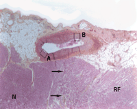
Higher magnification of Fig. 5. Arrows indicate the limit of radiofrequency effect area (RF) on epicardial myocardium. Left anterior descending artery enlarged at the origin of a diagonal artery. Half of the artery was in contact with the area exposed to RF (HES stain, ×25). A is a part of the artery not exposed to RF and B a part exposed to RF effect. N is the normal myocardium.

4 Discussion
The main findings of this study are that application of RF 1 mm from the coronary artery causes a significant decrease of endothelium-independent contraction to KCl and a significant decrease of endothelium-dependent relaxation to BK. There was no significant differences with the control group when RF energy was applied at 5 and 10 mm from the coronary artery. Histologic examinations confirmed the functional findings showing disruption of the vascular wall up to 5 mm from the RF application site.
The size of lesions induced by RF energy depends of various factors, anatomical as well as type of devices and procedure (power, duration, temperature) used. In the present study, in which RF was applied on the ventricular wall, which is thicker than the atrium, RF lesions penetrated deeply (5 mm) into the myocardium. The parameter used (20 W power and 20 s duration) were less aggressive than the settings used in previous reports [12–14] in which the size of the lesion increases with the duration of the ablation [15] even when adjusted to the thickness of the myocardial wall. Wittkampf et al. have demonstrated, that the size of the RF lesion produced with a 2-mm large catheter tip electrode also depends on the power applied by the generator and that maximum lesion areas are achieved in less than 20 s [16]. However, in the present study, constant duration and power were selected for each lesion created to compare the effects of RF relative to its proximity to the coronary artery. The site of application was the same for all procedures on the LAD and RCA and only the distance of application from the artery varied. Furthermore, there is no difference between the endothelium-dependent reactivity of the LAD and RCA in swine as previously demonstrated in our laboratory (data not shown).
The better understanding of the role of the endothelium in regulation of the vascular tone and coagulation control has led to the knowledge that structural and functional integrity of the endothelial layer are fundamental to avoid spasms or thrombosis of the coronary artery during or after surgical procedures [9,10]. The proximity of the circumflex artery to lesions made during the Maze procedure, especially when a RF is applied between the pulmonary veins to the mitral annulus [11] may jeopardize its integrity. As observed in the present experiments, when the coronary artery is involved in the necrotic process, both muscular and endothelial layers bear morphological lesions with edema, intimal disruption, exposure of the subendothelial layer, and muscular necrosis. These findings have functional implications including the decrease of muscular dependent contraction to KCl and decrease or suppression of endothelium-dependent relaxation in arteries located 1 mm away from the site of RF application and not at 5 mm, demonstrating that the section of the artery located outside the zone of RF effect maintains its functionality. The decreased contractions at 1 mm reflect morphological alterations of vascular smooth muscle cells described above but the level of contraction obtained was sufficient after exposure to PGF2α to study endothelium-dependent relaxations, which was also significantly altered.
4.1 Limitations
The first limitation of this study concerns creation of RF lesions in normal porcine ventricular myocardium with normal coronary arteries. However, patients treated with RF for chronic atrial fibrillation may also be free of coronary disease. Secondly, in clinical practice, RF is usually applied on the atrial endocardium rather than on the ventricular epicardium. The ventricle was chosen as the site of radiofrequency ablation to allow perfect visualization of the coronary arteries and a reproducible accuracy of the measurement of the distance between RF application in relation to the artery. The choice of ventricular wall also allowed accurate assessment of the maximum depth of extension of the lesions. Since application of RF ablation in these experiments involved the ventricular myocardium where the thermal spread is very limited, compared with the atrial wall, the usual site of radiofrequency ablation in the surgical practice, which has a great propensity for thermal spread, consequently, the present study is probably an underestimation of the actual risk of coronary lesions induced by radiofrequency during clinical procedures. The lesions induced in this beating heart model may not reflect exactly those made in the atrium during cardiac surgery in humans in which lesions are performed on an arrested heart after cardioplegia administration and at lower myocardial temperatures in case of hypothermic cardioplegia, which could potentially reduce the size of the lesions. Obviously this would not be the case when normothermic or tepid cardioplegia are used. In preliminary experiments of endocardial left atrial RF application performed under cardiopulmonary bypass and cardioplegic arrest there was no evidence of endothelial dysfunction of the circumflex artery, when compared to LAD and RCA used as controls (data not shown). Direct anatomical measurements of the distance between the endocardium and the circumflex artery varied from 6±1 mm in porcine explanted hearts, which could explain the lack of endothelial dysfunction observed in this set of experiments. The circumflex artery was used as control in organ chamber experiments with epicardial application of RF because its limited length in swine precluding its use for the study of the distance-lesion relationship.
4.2 Conclusion
Radiofrequency ablation with a 20 W power, 60 °C temperature and 20 s duration for 2 cm ablation induces coronary endothelial dysfunction associated with morphological damages when applied less than 5 mm from the coronary artery, in a beating heart porcine model. As the extent and depth of RF-induced lesions may vary depending on anatomical characteristics, the presence of blood, the probe temperature, the power, and duration of application, this study shows that a RF ablation can induce endothelial dysfunction and structural lesions of the coronary endothelium. Caution must be exerted during RF application in the left atrium due to the proximity of the circumflex artery and during right atrial RF application due to the proximity of the RCA. Techniques avoiding creation of lesions at these critical sites, especially near the mitral annulus, should be favored to prevent arterial damage and clinical complications related to coronary artery injury.
This work was supported by the Montreal Heart Institute Research Foundation and Surgical Department of ‘Université de Montréal’. We wish to thank Annik Fortier from the Department of Biostatistics and Diane Campeau for the preparation of the manuscript. Dr. Perrault is a scholar (Junior 2) of ‘Fonds de la recherche en santé du Québec’ (FRSQ).
Dr F. Mohr (Leipzig, Germany): I truly enjoyed this experimental study because it confirms the clinical findings. In a series of 380 radiofrequency ablation for A-fib surgery we have had two patients who had a circumflex artery narrowing or stenosis post ablation, and it led us to change the area of application. You showed your connection to the mitral valve at the P2 level, and after it occurred in case No. 200 or 200 something, we switched very high up to P3. The question is whether other thermal energy sources may cause the same injury. In a previous study it has been avoided to connect to the mitral valve. Do you think other energy sources would have less impact than radiofrequency?
Dr Demaria: I think all energy would be very dangerous for the artery, and probably cryotherapy should be a good option for that when we are obliged to do ablation near the circumflex artery. We do that in clinical practice.
