-
PDF
- Split View
-
Views
-
Cite
Cite
Kalliopi Athanassiadi, M. Gerazounis, Nikolitsa Kalantzi, P. Kazakidis, Anastasia Fakou, D. Kourousis, Primary traumatic pulmonary pseudocysts: a rare entity, European Journal of Cardio-Thoracic Surgery, Volume 23, Issue 1, January 2003, Pages 43–45, https://doi.org/10.1016/S1010-7940(02)00653-X
Close - Share Icon Share
Abstract
Objective: Pulmonary contusion is the usual manifestation of lung parenchymal injury after blunt chest trauma. With deceleration these parenchymal lacerations can result in cavities known as primary traumatic pulmonary pseudocysts (TPPC). We present our experience in treating this rare entity. Material: From 1989 trough 1999, 14 young patients, 11 male and three female ranging in age between 13 and 24 years were treated for primary TPPC in our department. Blunt chest injuries resulting from traffic accidents were the causes in all our cases. The main symptoms were pain, hemoptysis and dyspnea not associated with severe hypoxemia. The cavitary lesion was apparent in chest radiographs, but the imaging modality of choice was the computed tomography. Results: Multisystem injury was present in 7 of them. Two of our patients required ICU facilities but none needed mechanical ventilation. Hemopneumothorax was present in two cases, whereas pneumothorax in four drained by tube thoracostomy. The hospital stay ranged between 9 and 23 days. Contraction and complete radiological resolution of the PPC needed a follow up of 6–11 weeks. Conclusions: (1) Primary traumatic pulmonary pseudocysts are benign lesions secondary to blunt chest trauma needing only conservative treatment unless complications arise, such as hemo- or pneumothorax or infection of the cavitary lesion. (2) Computed tomography is a really sensitive method for early detection of the lesion while plain roentgenograms are sufficient for the follow up.
1 Introduction
The spectrum of lung parenchymal injury following blunt chest trauma ranges from simple contusions to lacerations [1]. A pulmonary contusion is characterized by hemorrhagic edema of the alveolar and interstitial spaces and usually resolves unless complicated by infection or acute respiratory distress syndrome (ARDS), while the term laceration is used for extensive visceral pleural disruption complicated by persistent bleeding or major air leak [1–3]. With rapid deceleration, however, parenchymal lacerations can result in cavities best termed ‘traumatic pulmonary pseudocyst’ (TPPC). The development of TPPC is a rare occurrence most often seen in children and young adults, in whom the thorax is elastic, the visceral pleura intact and the parenchyma easily injured, when a large pressure gradient is generated between a proximal obstruction and multiple hyperinflated alveoli [1–3].
We present our modest experience in treating this rare entity.
2 Material
From 1989 through 1999, TPPC were seen in 14 young patients, a number that represents the 2.1% of the pulmonary parenchymal injuries and the 0.1% of all chest injuries treated in the Thoracic Surgery department of the General Hospital of Piraeus, which is also a Level I trauma center. There were 11 male and three female ranging in age between 13 and 24 years. Blunt chest injury resulting from traffic accidents was the cause in all our cases.
The main symptoms were pain, hemoptysis and dyspnea. Hemoptysis although never severe and life threatening was noted in all our patients when they were encouraged to cough. Dyspnea was not associated with severe hypoxemia (the arterial oxygen tension ranged between 72 and 85 mmHg) and was often attributed to the pain the patient complained for.
The location of the TPPC was in the majority of cases (n=12) in the right lung.
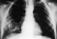
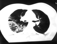
The cavitary lesion was apparent in chest radiographs. An air-fluid level was seen and the surrounding lung often showed consolidation. In 9 of our patients the TPP was detectable on the chest X-ray taken on the day of injury (Fig. 1) . Computed tomography (CT) was particularly useful in the demonstration and differential diagnosis of pseudocysts (Fig. 2) . All of them were depicted clearly not obscured by the pulmonary contusion shadow. Even in the five cases where the TPPC was not detectable on the chest X-ray because of coexisting pulmonary contusion or pulmonary hematoma, CT was useful for early diagnosis. In one case the cavitary lesion was accompanied by collapse of the middle lobe.

Chest roentgenogram at the time of admission showing the cystic lesion in the right lung along with contusion.

CT scan displaying infiltrates right-sided cystic lesions with an air-fluid level.
No haematological or biochemical abnormalities were revealed.
3 Results
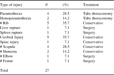
Multisystem injury was present in 7 of our patients. They included rib fractures, spleen and liver rupture, head injury, fractures of the scapula, etc. Hemopneumothorax was present in two cases, whereas pneumothorax in 4, drained by tube thoracostomy (Table 1) . Two of our patients required ICU facilities but none needed mechanical ventilation.

The hospital stay ranged between 9 and 23 days, while for those sustained only thoracic injuries the hospital stay decreased to 9–14 days. No secondary infection of the cyst was observed. Contraction and complete resolution of the TPPC needed a follow up of 6–11 weeks with chest X-ray. It showed appreciable reduction in size of the cavitating lesion and resolution of the surrounding consolidation.
4 Comments
Blunt chest trauma frequently produces pulmonary contusions, hematomas or effusions, but rarely leads to the appearance of a cystic lesion [1–5]. Since first described by Fallon in 1940 [6], these cystic lesions are stated in the literature with different names, such as cavitary pulmonary lesions, pseudocysts, pseudocystic hematomas, traumatic cysts, or traumatic lung cavities [1,3,4]. The authors agree with Santos and Mahendra [7] that the term ‘pseudocyst’ is correct, since the injured area does not include epithelium or bronchial wall elements as it is in a true cyst, nor it is a result of an infectious process as in a pneumatocele presented in children. The pathology of the cysts in surgically treated patients showed haemosiderin laden macrophages and fibrosis of the surrounding tissue, while their wall seemed to be derived from the interlobar connective tissue [1].
These lesions are either a direct result of the injury itself (primary pseudocyst) or can develop after resolution of a pulmonary hematoma (secondary pseudocyst) [8]. In our series, all cysts were primary since they were detected with 48 h from the accident.
The pathogenesis of TPPC is controversial. In an experimental rabbit study Lau et al. [9] demonstrated that impact velocity as well as chest wall displacement determines the severity and distribution of parenchymal injury. A high velocity causes alveolar injury, while a low velocity high displacement impact produces central parenchymal and major bronchial disruption. All our patients were involved in high-speed accidents, a fact that justifies both, the development and the relative peripheral location of TPPC. Eight-five percent of patients with traumatic lung pseudocysts were reported to be younger than 30 years [1]. The higher incidence in young adults may result from the greater compliance of the immature chest wall, permitting larger transmission of the force of impact to the parenchyma [2,10–12].
Hemoptysis, chest pain and cough were the symptoms the patients complained for and which were attributable to the pulmonary parenchymal injury and not to the TPPC itself. On the chest radiograph an air-fluid level is usually seen and the surrounding lung often shows consolidation due to pulmonary contusion [4]. Computed tomography can define more precisely the location and size of the cyst and may also be a useful tool for early detection and differential diagnosis [1,2,10]. Radiological resolution usually occurs within 2–3 months [3–5,10–13], as it proved to be in our series too.
Treatment is typically limited to symptomatic relief, as resolution usually occurs within a few weeks. Physiotherapy can be very useful for all patients sustaining blunt chest trauma, while bronchial ‘toilette’ is only suggested for the ones under mechanical ventilation. Generally, neither antibiotics nor surgical intervention is required unless complications arise [1–4]. Ribet [14] suggested that evolution of such localized lung injuries depends on three factors, the quality of surrounding tissue and the extent of contusion, the bronchiolar obstruction with rupture, hemorrhage or edema and finally infection. Infection of these pseudocysts is reported very rarely. Caroll et al. [15] stressed that infected traumatic PPC can be life threatening, because they may not respond like typical lung abscesses to appropriate antibiotic management. Early exploratory thoracotomy should be considered in those patients with prolonged fever and pulmonary deterioration. Successful operative debridement and drainage of the involved lung and pleural space may be life saving from septic complications. In conclusion, we would like to summarize.
Traumatic pulmonary pseudocysts are benign lesions secondary to blunt chest trauma, which need only conservative treatment unless complications such as hemo- or pneumothorax or infection of the cavitary lesion arise. The cavitary lesions usually diminish in size and resolve within or 2 or 3 months.
CT scan is a really sensitive method for early detection of the lesion while plain roentgenograms are sufficient for the follow up.
References
- computed tomography
- chest injury trauma blunt
- hypoxemia
- dyspnea
- lung
- hemoptysis
- traffic accidents
- deceleration
- follow-up
- hemopneumothorax
- intensive care unit
- lacerations
- pain
- chest x-ray
- thoracic injuries
- infections
- diagnostic imaging
- pneumothorax
- pseudocyst
- contusion of lung
- mechanical ventilation
- tube thoracostomy
- trough concentration
- conservative treatment
- early diagnosis


