


Abstract
This report describes the formation of a true aneurysm 15 years after an internal mammary artery patch aortoplasty of an aortic coarctation. A true aneurysm was confirmed on histology. To our knowledge, this is the first report of a case with such a complication.
1 Introduction
In 1972, Moor et al. [1] described a technique of patch aortoplasty for coarctation using the internal thoracic artery. Experience with this technique was reported with no aneurysm formation or re-coarctation [2]. The case here in is of an aneurysm formation following an internal thoracic artery patch repair of a coarctation after 15 years.
2 Case report
This case is that of a 2-year-old male who presented to his general practitioner with recurrent chest infections. He had mild pigeon chest deformity on inspection with an accentuated second heart sound and a loud systolic murmur. His blood pressure was measured at 160/110 and there was mild radiofemoral delay. Echocardiography demonstrated coarctation of the aorta and a bicuspid aortic valve causing minimal left ventricular outflow tract obstruction.
On 22 April 1985, the coarctation was repaired by patch aortoplasty utilizing the left internal mammary artery. His postoperative recovery was satisfactory. Persistent postoperative hypertension was treated initially with propranolol, later changed to atenolol.
2.1 Follow-up
Echocardiograms in 1986 and 1987 were satisfactory. A magnetic resonance image (MRI) in 1988 suggested mild re-stenosis. Cardiac catheterization demonstrated a gradient of 10 mmHg and indicated early aneurysm formation at the site of the previous repair. Repeated MRI demonstrated no new change until 1993 but thereafter there was a progressive increase in the size of the aneurysm. This measured 25 mm in 1998 and 29 mm 1 year later (Fig. 1) .
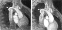
The increase in size of the aneurysm (white arrowhead) from 1998 (A) to 1999 (B).
The aneurysm was resected in February 2001 and the aorta reconstructed with an 18-mm Dacron interposition graft. The histology of the internal mammary artery patch, which was resected, showed focal loss of media with fibrous replacement, which had resulted in aneurysmal dilation (Fig. 2) .
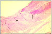
Histology showing loss of media (black arrow) and fibrous replacement (black asterisk).
3 Discussion
The advantage of patch aortoplasty over other standard techniques for repair of coarctation of aorta is that it involves minimal mobilization of the aorta and its collateral vessels. In addition, where the subclavian artery has been used in neonates and infants, the potential for growth has resulted in a low incidence of re-stenosis. In older patients, patch aortoplasty using a prosthetic material has resulted in a high incidence of aneurysm formation.
Although the incidence of aneurysm formation may be influenced by the choice of prosthetic material, it has been postulated that the cause of aneurysm formation is a result of the differing tensile characteristics of the patch and the aortic wall. In theory, by using the internal mammary artery, the influence of this causative factor is reduced. There have been several reports related to the use of the internal mammary artery for patch aortoplasty as a free graft [1–3] and others have proposed the use of a pedicle graft to retain the growth potential and integrity of the internal mammary artery [4]. Aneurysm formation of the internal mammary artery patch has not previously been described although there have been a number of aneurysmal dilations at the site of repair following subclavian flap aortoplasty [5].
The internal thoracic artery being a muscular artery and shows little of the elastic qualities of an aorta. Also, the lack of growth of the graft resulted in fracturing and loss of the media from the stretch induced by the higher blood pressure over time. The subsequent fibrous replacement resulted in a weaker patch, which again was not suited to the functional requirement of the aorta. This, we feel, resulted in the aneurysmal dilation of the patch over the years.
This case therefore calls into question the supposed theoretical advantage from the use of the internal thoracic artery as a patch provider for such a repair. The case also re-emphasizes the importance of continued close follow-up of all patients who have had repair of coarctation regardless of the particular method of repair.
