


Abstract
A 46-year-old man with Marfan syndrome was admitted for repair of annuloaortic etasia and funnel chest. Before median sternotomy, seven transverse skin incisions were made for resection of deformed ribs. The convex portions at the costochondral junctions of the right 4∼7th and left 5∼7th ribs were removed. Thereafter, the conventional median sternotomy was safely performed. Aortic root was replaced. After weaning from the cardiopulmonary bypass, the redundant distal end of the sternum was resected, fractured sites of the concave sternum were straightened and secured with wire fixation, and the split sternum was sutured with wires in an ordinary fashion.
1 Introduction
Marfan syndrome is an inherited connective tissue disease that affects the cardiovascular system, skeleton such as funnel chest, and others. Ravitch type procedures or sternal turnover can be chosen for patients with funnel chest based on cosmetic, psychological, or physiological reasons. Funnel chest is an isolated chest wall abnormality but can exist with cardiac diseases. Especially, in Marfan syndrome, associations between funnel chest and cardiovascular diseases are well described. Cardiac surgery requires a good operative field, while the deformed thorax may prevent surgeons from getting it. When the deformed sternum is removed for sternal turnover, the free bone graft is exposed to ischemia during cardiac surgery. We report a Marfan patient undergoing a repair of severely deformed thorax without dissection of the deformed thorax associated with simultaneous repair of annuloaortic ectasia.
2 Technique
A 46-year-old man with Marfan syndrome was referred to our hospital for repair of annuloaortic etasia and funnel chest. On admission, funnel chest, vertebral scoliosis, and hump back were observed. In addition, a diastolic murmur of Levine II was also audible. Chest computed tomography (CT) showed severely excavation of the sternum and kyphotic change of the back (Fig. 1A) . Aortography and echocardiography revealed annuloaortic ectasia with aortic valve regurgitation of grade IV. The patient consented to undergo simultaneous repairs of the cardiovascular lesion and deformed thorax.
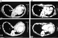
Pre- and post-operative chest computed tomographic findings. (A) Preoperatively, a severe excavation of the anterior chest wall and displacement of the mediastinum are recognized. (B) Postoperatively, the deformed wall is repaired, while the mediastinum remains displaced.
Before median sternotomy, seven transverse skin incisions were made for resection of deformed portions of the ribs (Fig. 2A) . The convex portions of 2∼3 cm in length at the costochondral junctions were removed, while the intercostal neurovasculartures were remained intact. The right 4∼7th and left 5∼7th ribs were severed there. Thereafter the sternum could be elevated, and the conventional median sternotomy was safely performed. Application of a sternal retractor caused transverse fractures at the two sites of each divided sternum (Fig. 2B). Under hypothermic cardiopulmonary bypass, aortic root was replaced with a Hemashield Gold (Meadox Medical Inc., Oakland, NJ) of 28 mm and Bicarbon valve (Sorin Biomedica Cardio, Saluggia, Italy) of 23 mm. After weaning from the cardiopulmonary bypass, the redundant distal end of the sternum was resected (Fig. 2C), fractured sites of the concave sternum were straightened and secured with wire fixation (Fig. 2D), and the split sternum was sutured with wires in an ordinary fashion. The severed ribs were not approximated. Two week mechanical ventilation was programmed to stabilize the surgically intervened thorax. Thereafter, the patient's chest wall was stable with an excellent cosmetic appearance. Chest CT showed an acceptable correction of deformed thorax 2 months after operation (Fig. 1B). The patient is doing well 1 year and a half after operation with cosmetic improvement of the thorax, and he is satisfied with the surgical results of the thoracic deformity.
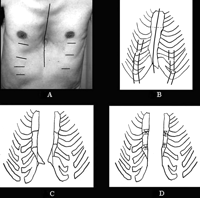
Operative procedures of funnel chest. (A) Skin incisions are shown. Seven incisions were made for resection of the convex costochondral junctions. After the resection, an ordinary median sternotomy was performed. (B) The right 4∼7th and left 5∼7th deformed ribs of 2∼3 cm in length are resected. (C) Application of a sternal retractor caused transverse fractures at the two sites of each divided sternum. The convex xyphoid process obstructed aligning the straightened sternum. (D) The deformed sawed sternum is repaired, being straightened by overlapping each fractured end and securing by wire fixation following resection of the redundant distal ends of the sternum. The sternum is sutured with wires in a usual fashion.
3 Comment
For simultaneous repairs of both deformed thorax such as funnel chest and cardiac diseases, how to repair the thorax is the key issue. Sternal ischemia for long hours may cause sternal complications. Shamberger and colleagues stated that simultaneous cardiac and pectus excavatum repair should be avoided [1]. However, some one-stage procedures have been anecdotally reported [2–6].
Sternal elevation such as a Ravitch type procedure requires dissection around the sternum and associated ribs from the pectoral muscles with bilateral subperichondrial resection of the deformed costal cartilages and sternal osteotomy resecting a wedge of the anterior cortex and fracturing the posterior cortex, resulting anterior displacement of the sternum [1,7,8]. To get a good operative field, harvesting of the sternal graft may precede cardiac operation when simultaneous repairs of the sternal deformity and cardiac lesions. The sternal elevation may be adequate in cases of atrial or ventricular septal defects because of the short ischemia time of the sternal graft. However, repairs such as aortic root replacement require longer ischemia time for the free graft than those in case of atrial or ventricular septal defect. Sternal elevation per se can cause sternal graft failures such as necrosis, infection, bleeding, and others [1,7]. The longer the ischemia time is, the higher the possibility of the complications is. Minimizing sternal dissection or devascularization can be the key for simultaneous repairs of both lesions.
Our procedure does not require the sternal osteotomy for repair of the deformed sternum, nor the pectoral muscle mobilization for resection of the deformed ribs. We make several small skin incisions to resect bilateral deformed costochondral junctions with intact intercostals neurovasculartures, preserving the perichondral and periosteal sheath of costal cartilages and ribs. Thus, the sternum is not exposed to ischemia, and the dissection of the graft can be minimized. Through additional conventional medial sternotomy and simple sternal plasty with wires, the deformed thorax could be cosmetically repaired. We scheduled 2-week respiratory assist to get possible sternal instability. Up to 1 year and a half follow-up after operation, the cosmetic appearance is quite excellent without sternal complications.
When two-stage repairs for deformed thoraxes and cardiac lesions are chosen, some surgical interventions such as endoscopic or conventional procedures for the former may precede repairs of the latter. However, sternal devascularization or ischemia should be minimized in case of simultaneous repair avoiding sternal complications. A long-term follow-up and additional experiences are necessary to confirm the safety and effectiveness of our method for repair of funnel chest.
