-
PDF
- Split View
-
Views
-
Cite
Cite
Antonio Ríos Zambudio, Juan Torres Lanzas, María José Roca Calvo, Pedro J. Galindo Fernández, Pascual Parrilla Paricio, Non-neoplastic mediastinal cysts, European Journal of Cardio-Thoracic Surgery, Volume 22, Issue 5, November 2002, Pages 712–716, https://doi.org/10.1016/S1010-7940(02)00484-0
Close - Share Icon Share
Abstract
Objective: The non-neoplastic mediastinal cysts (NNMCs) form a group of uncommon benign lesions of a congenital origin. The significant controversy regarding these cysts is whether to manage with observation or surgical resection. The aim of this study is to analyse the utility of thoracic computed axial tomography (CT) in imaging diagnosis of the NNMCs and the results of surgery in these lesions. Patients and methods: Twenty NNMCs underwent surgery between 1980 and 2000. The preoperative study of mediastinal cystic masses includes a complete blood test, chest radiography (CR) and, for the last 15 years, a thoracic CT and/or nuclear magnetic resonance. All the patients underwent surgery in our thoracic surgery department and were reviewed in outpatients at 1 month, 6 months, 1 year and biannually thereafter. The form of manifestation, clinical features, imaging techniques, surgical operation, morbidity, mortality and follow-up are analysed. Results: Ten corresponded to bronchogenic cysts, the most common symptom of which was chest pain. CR showed a mass in the anterior-superior mediastinum in nine cases, and CT (five cases) revealed a cystic tumour in the anterior mediastinum. All were removed surgically, with three patients presenting with mild complications. Seven corresponded to pleuro-pericardial cysts, four being asymptomatic. CR showed a right paracardial mediastinal tumour, which was confirmed by CT (four cases). All were removed surgically, with two patients presenting with mild complications. Three corresponded to enteric cysts. CR showed a tumour in the posterior mediastinum, with CT confirming its cystic nature (two cases). Excision of the cyst was done in all cases, which corresponded to duplication cysts: two oesophageal and one gastric. All the patients are asymptomatic and recurrence-free after a follow-up of 11±10 years. Conclusions: NNMCs are benign lesions in which the lesions in which the surgery can be done with a low morbidity and mortality rate, enables us to rule out malignancy and offers a definitive cure. Actually the thoracic CT permit a correct diagnosis pre-surgery in function of the radiologic characterisation and topography.
1 Introduction
The mediastinum is an anatomical space in which a number of both neoplastic and non-neoplastic pathologies appear. Within this group, non-neoplastic mediastinal cysts (NNMCs) form a group of uncommon benign lesions of a congenital origin, due to the abnormal development of the tracheobronchial tree and/or primitive intestine. They are generally asymptomatic, unless they attain a large size and cause compressive symptoms, which is often a incidental radiologic finding [1,2]. They are particularly significant because of the difficulty in making a differential diagnosis, as they can simulate multiple lesions, both benign and malignant. Currently, with the improvement in non-invasive techniques, a pre-surgical diagnosis can be made in a high percentage of cases, although it is relatively common for surgery to be indicated to establish the definitive diagnosis. One controversial aspect of these tumours is the treatment to give, which ranges from observation to surgical resection, without there being a consensus on the best therapeutic option [1,2].
The aim of this study is to analyse the utility of thoracic computed axial tomography (CT) in imaging diagnosis of the non-neoplastic mediastinal cysts and the results of surgery in these lesions.
2 Patients and methods
Between 1980 and 2000, 20 patients underwent surgery in our department with a definitive anatomico-pathological diagnosis of benign mediastinal cyst, this being understood as one of the following: (1) bronchogenic cyst; (2) pleuro-pericardial cyst; and (3) enteric or duplication cyst. The cystic lymphangioma, cystic teratomas and cystic thymomas were excluded because they are neoplastic pathologies and occasionally can be cyst. The patients data were obtained from a retrospective review.
The mean age at presentation was 42±13 years, and most of the patients were women (13 cases). The preoperative study of mediastinal cystic masses includes a complete blood test, chest radiography and, for the last 15 years, a thoracic CT scan. In the last 3 years the preoperative study has come to include, in cases with dubious diagnoses, nuclear magnetic resonance (MRI).
All the patients underwent surgery in our thoracic surgery department and were reviewed in outpatients at 1 month, 6 months, 1 year and biannually thereafter.
We analyse the patient sex and age, form of manifestation, clinical features, results of the imaging techniques (chest radiography, thoracic CT scan, and/or thoracic MRI), localisation of cyst in mediastinum, size, type of surgical operation performed, morbidity and mortality rates during the immediate postoperative period (1st month), follow-up and cases of recurrence.
3 Results
Of the 20 patients ten corresponded to bronchogenic cysts, seven to pleuro-pericardial cysts and three to enteric cysts (two oesophageal duplication and one of a gastric origin).
3.1 Bronchogenic cysts (n=10)
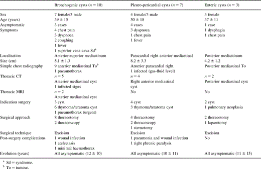
The mean age at presentation was 39±15 years; seven cases were women. The most common symptom was thoracic pain in four cases followed by dyspnoea in three. Blood tests were normal in all the patients, except in one case, who presented with chronic anaemia which had been under treatment with iron for a year. Simple chest radiography revealed a mass effect in the anterior-superior mediastinum in nine cases. CT was done in five patients and revealed a cystic tumour in the anterior mediastinum. MRI was performed in the two most recent cases, which confirmed the presence of the cyst (Table 1) .

All the patients underwent surgery, the approach being a thoracotomy in eight cases and a thoracoscopy in the rest. Only three patients had a preoperative diagnosis of benign mediastinal cyst. The main diagnostic doubt, especially in the pre-CT era, was with thymoma and cystic teratoma in six patients (Table 2) . All the cases had excision of the lesion. The mean size of cyst was 5.1±1.3 cm. During the postoperative period three patients presented with mild complications (one wound infection, one minimal haemothorax and one atelectasis). After a follow-up averaging 12±10 years no recurrence has been noted, and all the patients are asymptomatic.

3.2 Pleuro-pericardial cysts (n=7)
The mean age at presentation was 50±18 years, and four were men. Four patients were asymptomatic, the cyst being a casual radiological finding. The other three presented with dyspnoea. Blood tests were normal in all the patients. Simple chest radiography in all cases revealed a right paracardial mediastinal tumour, which was confirmed by CT in the four cases in which it was done (Table 1).
All the patients underwent surgery: four with thoracotomy, one with sternotomy, and the rest with video-thoracoscopic surgery. In the four cases receiving CT the preoperative diagnosis was a pleuro-pericardial cyst; in the other three there was a diagnostic doubt with thymoma and cystic teratoma (Table 2). The cyst was removed in all the cases. The mean size of cyst was 8.2±3.3 cm. Two cases presented postoperative morbidity (one pneumonia and wound infection; and one asymptomatic right phrenic paralysis). After a follow-up averaging 10±11 years all the patients are asymptomatic.
3.3 Enteric or duplication cysts (n=3)
The mean age at presentation was 37±11 years, and all were women. One patient was clinically asymptomatic, another presented with thoracic pain, and the third dysphagia. The blood test was normal in two cases, the other revealing microcytic chronic anaemia and hypochromia. A CT scan was done in the two more recent cases, which confirmed the cyst preoperatively (Table 2). The third patient underwent surgery diagnosed with pulmonary neoplasia. The approach was thoracotomy in two patients and a midline laparotomy in the other. Excision of the cyst was performed in all cases. Pathological anatomy informed of oesophageal duplication cyst in two patients and a triple gastric duplication cyst and leiomyoma of the oesophagus in the third. The mean size of cyst was 4.2±1.2 cm There were no complications in the postoperative period. The three patients are asymptomatic 2, 3 and 28 years after surgery (Table 1).
4 Discussion
NNMCs are generally congenital and form around the 6th week of gestation, although pleuro-pericardial cysts can occasionally be acquired [2]. They account for approximately 20% of primary mediastinal lesions, the most common being bronchogenic (50–60%), followed by pleuro-pericardial (20–30%) and, less frequently, enteric or duplication (7–15%) [2]. They may appear at any age, although they are more common in the 4th and 5th decades of life, generally detected in a routine radiological study [3], as observed in our patients. There are no differences between sexes except for neuro-enteric cysts, which are more common in females [2]. They may be located in any part of the mediastinum, although bronchogenic cysts have a preference for the mid and superior mediastinum [3], pleuro-pericardial cysts for the right anterior cardiophrenic angle [4], and enteric cysts for the posterior mediastinum [2].
NNMCs in adults usually begin as a incidental radiological finding in asymptomatic patients [5]. They may present a variety of symptoms, particularly coughing and chest pain, which are generally caused by the compression of neighbouring structures [2]. In the absence of complications, clinical features depend on the site of the cyst: paratracheal and carinal cysts may lead to tracheobronchial compression, causing coughing, wheezing, dyspnoea and stridor; para-oesophageal cysts can cause dysphagia, regurgitation and abdominal pain [1,2,5,6]. Neuro-enteric cysts with intraspinal spread can appear with neurological symptoms [7]. The most serious complication, though fortunately rare, is malignant degeneration [5].
Chest radiography usually shows a well-delimited, homogeneous, spherical mediastinal image. Bronchogenic cysts are typically paratracheal or subcarinal, the pleuro-pericardial adhere to the heart and diaphragm, and the duplication are posterior [4,8]. When they are infected or communicate with an airway or digestive tract, an air bubble is produced inside the cyst [5], as observed in some of our patients.
CT has increased the diagnostic performance of non-invasive imaging techniques. It shows a well-defined spherical cystic lesion with a watery content of attenuated intensity and delimits its connection with neighbouring structures, especially the oesophagus and airway [2,8]. In the pleuro-pericardial cysts the wall is imperceptible and is located paracardially [2]. When there is communication with the tracheobronchial tree, a gas-fluid level is seen in the cyst [9]. Currently MRI seems to provide a better definition of the cyst and its connection with neighbouring structures than CT, showing low-signal intensity images in T1 sequence and bright-signal intensity images in T2 [8,10]. Enteric cysts form during early embryogenesis when the anterior intestine and notochord are close, which is why abnormalities of the vertebral column are usually associated [2]. For this reason MRI should be done in these patients to exclude the intraspinal spread of posterior mediastinal cysts. Other tests are useful for ruling out complications; for instance, gastro-intestinal endoscopy and bronchoscopy rule out communication of the cyst with the oesophagus or airway.
Clinical features and radiology may give us a clue to diagnosis, but no exploratory technique or clinical manifestation is characteristic, as several pathologies can be simulated [2,3]. A differential diagnosis must always be made with other cystic pathologies, especially as there are cystic lesions of a benign appearance that can mask malignant neoplastic lesions. In our hospital the CT is a exploration routine in this lesions in the last 15–17 years. Actually the radiologic characterisation is important, because one well-defined cyst with a watery content of attenuated intensity in a typical localisation is very suggestive of these benign lesions. Definitive diagnostic confirmation is anatomico-pathological. The histological study shows an epithelium-coated cyst, varying according to cyst type, inside which bronchial components may be found [3,5].
The treatment of choice is complete excision of the cyst, even in asymptomatic patients, in order to prevent complications and establish diagnosis [3,5,11]. The prognosis after complete excision is excellent [2,7], and the morbidity and mortality surgical rates are low.
Occasionally a conservative attitude has been considered, with a clinical and radiological follow-up without surgery, especially with pleuro-pericardial cysts [2,9]; however, this is a controversial subject. There are those who recommend conservative treatment as it avoids surgical morbidity and mortality, and there have even been reports of spontaneous resolution of the cyst [12]. Those against an expectant attitude show that resection carries little morbidity and mortality, and many of the patients that do not receive surgery at the time develop symptoms [13] related to cyst growth, which means that an operation then will involve a higher morbidity and mortality rate, together with a risk of malignancy and development of complications [14]. What is indeed clear is that surgical excision is a must when some of the following criteria are met [5]: (1) symptomatic cyst; (2) suspected malignancy; (3) cyst infection; (4) tracheal compression; (5) progressive growth; (6) presence in children; or (7) atypical location or characteristics. This paper present a surgical series. We have not untreated mediastinal cysts. Only two cases with a cyst lesion in anterior paracardial mediastinum with a size of 2 and 3 cm, suggest of pleuro-pericardial cysts, have not operated. The evolution is favourable. These cases are not included in the series because is a surgical study and the untreated cyst are only two.
In recent years the use of video-assisted thoracic surgery techniques has been demonstrated in several studies for NNMC resection, something also observed in the two cases in our series. Whilst more studies are needed to finally confirm its usefulness, it promises to be the standard approach for treatment, relegating the options of conservative therapy due to its low morbidity and mortality rates and quick recovery [15,16].
We can say, in conclusion, that NNMCs are benign lesions in which the surgery can be done with a low morbidity and mortality rate, enables us to rule out malignancy and offers a definitive cure. Actually the thoracic CT permit a correct diagnosis pre-surgery in function of the radiologic characterisation and topography.
References
- nuclear magnetic resonance
- pericardial sac
- chest pain
- computed tomography
- blood tests
- cancer
- bronchogenic cysts
- cysts
- follow-up
- mediastinal cyst
- mediastinal neoplasms
- outpatients
- preoperative care
- chest x-ray
- signs and symptoms
- surgical procedures, operative
- diagnosis
- diagnostic imaging
- mediastinum
- morbidity
- mortality
- neoplasms
- radiology specialty
- thoracic surgery procedures
- cystic neoplasms
- chest ct
- excision
- anterior mediastinum
- posterior mediastinum


