


Abstract
Objective: The Symmetry™ aortic connector creates proximal anastomoses of saphenous vein grafts using a nitinol implant. The device avoids partial clamping and thus possibly reduces neurologic complications. To evaluate graft patency, a single surgeon randomised study was performed in our institution. Methods: Seventy-seven patients were randomised either to automated proximal anastomoses (group I, n=39, 61 vein grafts connected using the aortic connector, 47 as single, 15 as sequential bypasses) or controls (group II, n=38, 62 proximal anastomoses handsewn, 46 as single, 16 as sequential bypasses). Ultrafast CT-scans were performed on postoperative day 5 in 34 patients of group I and 16 patients of group II to evaluate early graft patency. Intermediate term patency was evaluated with ultrafast CT-scan in 30 patients of group I (46 grafts) and 25 patients of group II (39 grafts) 1 year after the operation. Results: Two early graft occlusions were detected in group I (3.8%). In group II all evaluated grafts were patent 5 days after surgery. 11.4 months after surgery, seven out of 46 grafts were found occluded (15.2%) in group I. In the control group, only one occlusion was detected (2.6%). Conclusions: We observed a trend towards an increased occlusion rate 1 year after surgery with automated connector devices. For evaluation of long-term patency larger patient groups have to be evaluated. Other benefits of these devices have to be proven to promote their clinical application.
1 Introduction
Vascular anastomoses in coronary artery bypass grafting (CABG) are mainly performed by continuous sutures for proximal anastomoses. This not only means dependency on manual skills, experience and time, but requires the use of a side biting clamp with manipulation of the ascending aorta. This may cause consecutive detachment of debris and particulate embolization, possibly leading to neurological complications. Less traumatic techniques have been developed recently, and among other techniques several anastomotic devices are currently available. One of these devices is the Symmetry™ aortic connector (St Jude Medical Inc., Minneapolis, USA), creating proximal anastomoses with saphenous vein grafts using a nitinol implant. This allows a very fast connection of the saphenous vein graft to the aorta without partial clamping, possibly reducing trauma and handling of the aorta compared to the running suture technique [1]. This technique also may support minimally invasive CABG with the goal of reducing perioperative morbidity and therefore accelerate postoperative recovery. For off pump coronary surgery manipulation of the ascending aorta could be minimized. Despite the expected benefit, a certain amount of bypass occlusions or significant stenoses were described recently [2,3]. To evaluate short and mid term patency, we performed a single surgeon randomized study in our institution.
Ultrafast cardiac computed tomography (CT) as a noninvasive method for assessment of graft patency was suggested by the groups of Bateman (1987) and Stanford (1988), with a predictive accuracy of 96 and 92%, respectively. They concluded that technically adequate studies could be effectively performed in the majority of CABG patients. Despite a reasonable high temporal and spatial resolution of the scanner, limitations were seen in differentiation of obstructed and nonobstructed grafts. Details of proximal and distal anastomoses and the distal vasculature were not provided by recent CT scanners [4,5].
2 Materials and methods
2.1 Patients
We investigated 77 consecutive elective CABG patients who were prospectively randomized to either group I (aortic connector, n=39, 61 proximal anastomoses) or group II: handsewn, n=38, 62 proximal anastomoses) by a computer generated randomisation list. Inclusion criteria were age between 40 and 80, coronary heart disease with indication of aortocoronary bypass procedure, given informed consent, unimpaired renal function, no neurological history and no severe aortic calcification revealed by palpation by the surgeon during surgery.
Neither intraoperative or postoperative echocardiography nor graft flow measurement was performed routinely. Postoperative anti platelet therapy consisted in acetylsalicyl acid 100mg once a day for both groups starting postoperative day 1. The study was approved by our institutional review board and written informed consent was obtained from all patients. Unpaired t-test was used for detecting demographic or perioperative differences of the investigated groups. Chi-square test was applied to test for significance in occlusion rates between groups. There were no statistical significant differences between groups for demographic and perioperative data, see Table 1.
2.2 Surgical procedure
All operations were performed using extracorporal circulation (ECC). In group I we created 39 arterial bypasses to the LAD using the left internal mammary artery, six of those as jump grafts to a diagonal branch. Six right internal mammary grafts were performed either to the circumflex artery or to an intermediate branch, respectively. Among the 61 venous grafts connected to the aorta, 30 led to branches of the right coronary artery area, three of those were jump grafts. Twenty-eight venous grafts lead to the area of the circumflex artery, 13 as jump grafts. Two venous grafts led to a diagonal branch of the LAD and one to an intermediate branch. In total, we performed 128 distal anastomoses in group I (3.3 anastomoses/patient).
In group II, 27 of the 62 venous grafts sutured to the aorta led to the area of the right coronary artery, three of these as jump grafts. Twenty-eight grafts had their target in the circumflex area, 14 jump grafts were performed. Four were anastomosed to a diagonal branch and three to an intermediate branch. Additionally, we created 36 arterial bypasses with the left internal mammary artery to the left anterior descending artery, three of them as jump graft to a diagonal branch. One LITA graft was anastomosed to a posterolateral branch. Three right internal mammaria arteries were used as bypass graft to the circumflex artery (one sequential graft). The number of distal anastomoses amounted to 123 in group II (3.2 anastomoses/patient).
2.3 MDCT follow up
To evaluate patency we performed ultrafast computer tomography (MDCT) on 34 patients (53 proximal anastomoses) of group I and 16 patients (25 proximal anastomoses) of group II on the fifth postoperative day. In our early MDCT patency control we focused on the anastomatic connector group, 16 patients of group II served to validate our method. For evaluation of intermediate term patency we contacted all patients to get them for MDCT (see Fig. 1). One year (11.4±5 months) after the operation, 30 patients of group I (46 proximal anastomoses) and 25 patients of group II (39 proximal anastomoses) were investigated.
Chi-square test was applied to test for significance in occlusion rates between groups.
During the hospital stay two patients died. One in the aortic connector group died 3 days after the operation of perioperative myocardial infarction. The other one was randomized to group II and died three month after the operation without having left hospital. Between hospital discharge and second follow up one patient of group I died for unknown reasons 14 month postoperatively, and another patient of group II died of noncardiac disease. We were able to include five patients of group I and 15 patients of group II who had not participated in the immediate postoperative follow up.
The patient with the perioperatively occluded vein graft (group I) was not reinvestigated. So in group I out of 36 patients amenable to follow up 30 were seen, two had contraindications to contrast media and four withdrew their consent. In group II out of 36 patients 25 were seen four had contraindications and seven withdrew their consent (see Fig. 1).
2.4 Image acquisition and reconstruction
The CT scans at the early postoperative examination were performed using a multidetector 4-row spiral CT (SOMATOM Plus 4 Volume Zoom WIP-Version VA 20, Siemens Inc, Forchheim, Germany). At the 1 year follow up MDCT examinations were performed on a 16-row spiral CT (Sensation 16, Siemens Inc., Forchheim, Germany).
Image reconstruction in all patients was performed using retrospective ECG gating, a technique that allowed continuous image reconstruction from volume data sets during any phase of the cardiac cycle. Adaptive Cardiac Volume (ACV)® technique-served as reconstruction algorithm. Depending on the heart rate this technique combines two different reconstruction procedures: single-segment reconstruction (≦72bpm) and adaptive-segmental reconstruction (≫72bpm). Single-segment reconstruction requires data from only one rotation, adaptive-segmental reconstruction from up to two rotations for the reconstruction of each 1.0mm slice.
3 Results
Five days after the operation (follow up 1) we were able to investigate 50 patients, 34 patients of group I with 53 proximal anastomoses, and 16 patients of group II who were provided with 25 handsewn proximal anastomoses.
All in all we examined 55 patients at the 1 year follow up, 30 of the aortic connector group and 25 of the handsewn group. So we evaluated 85 proximal anastomoses at the 1 year follow up. Forty-six of these in the aortic connector group and 39 in the control group.
All patients were contacted and interviewed by us for the 1 year follow up. They were all in good clinical condition.
Of the 53 proximal anastomoses created with the aortic connector and evaluated at the perioperative follow up two were found to be occluded (3.8%). All other grafts were patent and had no stenosis at the proximal anastomotic site (see Fig. 2). But distal graft quality could not be examined for reasons of MDCT technique. There was no occlusion or proximal stenosis detected in the control group at the first follow up.
Of the 46 proximal anastomoses created with the aortic connector investigated after 1 year, we found seven grafts occluded (see Fig. 3). This calculates to an occlusion rate of 15.2%. In addition, another two grafts of this group presented with a 50% stenosis at the proximal anastomotic site (see Fig. 4). Out of the 39 hand sewn anastomoses of the control group examined at 1 year follow up we found one occlusion and no significant stenosis at the proximal anastomotic site. This gives an occlusion rate of 2.6%. All occlusions detected in mid term follow up were revealed in asymptomatic patients who did not need further interventional therapy. Chi-square test did not reveal statistical significance in occlusion rates between groups (P=0.5).
4 Discussion
Myocardial infarction due to graft occlusion was observed in one patient of our study group. Seven of 46 anastomoses created with the aortic connector and investigated at the mid term follow up after 1 year were found occluded. This represents an occlusion rate of 15.2% which does not exceed occlusion rates reported on large series of vein grafts in literature with 19% occlusion rate in the same period of time [8]. In the control group, only one of 39 hand sewn anastomoses was found to be occluded at 1 year follow up. Causes for early graft occlusion may be distal or proximal anastomotic problems, poor graft function or twisting of the vein graft or poor quality of the target vessel. Further investigations should also focus on the question if more aggressive anti platelet management could improve occlusion rates of the aortic connector group. Calafiore and Eckstein [9,10] reported possible advantages such as minimizing aortic manipulation but this has to be proven for clinical impact in further studies. In most patient cohorts, patency control was performed by coronary angiography. The group of Engelmann et al. confirmed in 1997 the limited effectiveness of CT for visualization of the distal anastomotic site and detection of possible graft stenoses by comparing CT scans to angiography. An accuracy of 96% for venous graft patency was reported by them. The same group had improved the accuracy of CT follow up to 100% regarding patency three years later [6]. By the use of ECG-triggering of conventional CT scanners visualization of proximal anastomoses was successfully demonstrated with evaluation of the anastomotic quality in 2000 [7]. Every new generation of CT scanners improves picture quality and thus reliability. In this study we were able to show that the use of MDCT is a reliable method for evaluation of graft patency and detection of proximal graft stenosis. We could show that the current technique of MDCT provides a less invasive and less expensive method for patency control and detection of proximal graft stenosis. This is of special interest because most patients are quite reluctant to undergo postoperative angiography. Due to motion artefacts the evaluation of distal anastomoses is still difficult, especially in the arrhythmic patient. Because of artefacts three proximal anastomoses could not be evaluated in satisfactory quality, however, graft patency could be proven. This problem may resolve with ongoing technical developments. Regarding distal anastomotic quality, coronary angiography provides more information than the CT scans of current technology and has the advantage that immediate intervention is possible in case of stenosis. For this reason, we performed coronary angiography in the patient presenting clinical signs of myocardial infarction. The other patient with a graft occlusion detected by MDCT also underwent coronary angiography, where our finding was confirmed, thus underscoring reliability of MDCT.
In our study group we observed a statistically nonsignificant trend towards increased occlusion rates in the aortic connector group. The percentage of occlusion in group I still meets occlusion rates described in the literature, whereas in our control group this percentage was lower. Statistical proof of this statement needs larger cohorts of patients. However, the Symmetry™ aortic connector may provide benefits if used in patients where aortic clamping is difficult or impossible, such as in minimally invasive procedures or in the case of calcified aortas. As it is our policy not to clamp or partially occlude a severely calcified aorta, these patients had to be excluded from this randomized trial, even if a partial calcification of the aorta may be a good indication for proximal anastomotic devices. A limitation of our trial is the fact that 29% of our patients initially included were not amenable for mid term follow up for reasons explained above.
We conclude, that occlusion rates of vein grafts anastomosed with the Symmetry™ aortic connector are equal to those described in the literature but showed a decreased intermediate term patency compared to our handsewn control group in this randomized study. Larger patient cohorts should be investigated to evaluate long term patency of grafts connected to the aorta using metal implants. MDCT provides reliable data on graft patency and quality of proximal anastomoses, even in the presence of nitinol implants.
Appendix A. Conference discussion
Dr W. Mohl (Vienna, Austria): Although you couldn't find out risk factors for the occlusion, do you have any impression whether there is a technological problem or is this the problem of how you put the graft in, the angle of the graft?
Dr Dietrich: It was just one of the perioperative findings that we had, for example, was there vascular disease or something like that, but the univariate analysis did not show any difference in any of these factors that could give us a hint for this occlusion rate.
Dr R. Gallo (Khamis Mushayt, Saudi Arabia): Could you please tell us which kind of antiplatelet aggregation do you use for the connectors? Are they the same as the other CABG?
Dr Dietrich: We applied aspirin, 100mg, as with the hand-sewn CABG group II. We did not change the therapy.
Dr Gallo: Just a short comment. Because we are starting to use it, we took this protocol from other groups, and they say that with the presence of nitinol, you can use clopidogrel, 75mg/day. Some groups say three months and other groups say six months. We are following the six-month protocol, and we are following by the same method like you, by ultrafast CT scan, and until now, we are just starting with only 27 patients, but we didn't find any occlusion.
Dr P. Tozzi (Lausanne, Switzerland): I have a question concerning the study design. You didn't measure the flow into the bypass, and I think it is difficult to compare the two groups if you have no information concerning the coronary runoff in the two groups. Could you comment?
Dr Dietrich: This is correct, we didn't measure the flow. With another study oncoming we would do though, that is correct. We have no data from this point, yes.
Dr R. Poston (Baltimore, MD, USA): First, you showed a CT angiogram with a Symmetry graft going to the right coronary that looked like it was kinked, because it did not come off into the right AV groove. Measuring flow might have pointed out that graft was taking an acute turn which compromised that particular graft that you showed us; presumably there were others in your series.
Second, the 15% graft thrombosis rate in your connector group is about on par with what you might see with early follow-up with most vein grafts. The less than expected 3% rate in the hand sewn group may have been influenced from the extensive loss to follow-up in that group. For instance, one guy died of an MI in that group prior to CT angio, who likely had an unmeasured graft thrombosis.
Mr V. Zamvar (Edinburgh, UK): My question is similar to the last one. Do you have any information on why there was such a large dropout in the two groups, particularly in the hand sewn group?
Dr Dietrich: No, we have no idea why in the hand sewn group we had such a small rate of occlusion or of stenosis. We couldn't discover any data giving us a idea why this difference occurred.
Dr P. Sergeant (Leuven, Belgium): You have no information on these patients? They were completely lost to follow-up?
Dr Dietrich: Patients lost to follow-up which we contacted but didn't want to come again for being examined, they were all well but just didn't want to go on with this.
Dr Sergeant: I have some residual problems with the study design. You have stated that this study is randomized. I see no power calculation. Has this study gone through the ethical committee of the hospital, because then the patients probably have agreed on this secondary evaluation in your study project?
Dr Dietrich: Well, they did agree to this second examination, but they just took back their agreement.
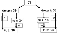
Recruitment for MDCT follow up (group I, aortic connector group; group II, Handsewn group: FU 1, follow up 1; FU 2, follow up 2).
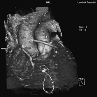
Patent aortic connector in three-dimensional reconstruction.
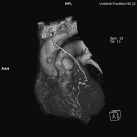
Occluded graft with aortic connector in three-dimensional reconstruction.
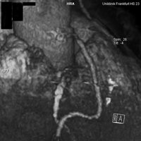
Fifty-percent stenosis of a graft with aortic connector in three-dimensional reconstruction.
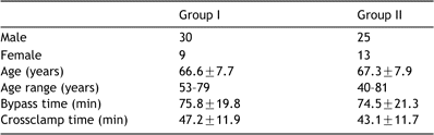
Demographic and perioperative data
Presented at the joint 17th Annual Meeting of the European Association for Cardio-thoracic Surgery and the 11th Annual Meeting of the European Society of Thoracic Surgeons, Vienna, Austria, October 12–15, 2003.
The study received financial support from St Jude Medical Inc.
