


Case description
A 73-year-old male patient with severe claudication was admitted to our hospital. Angiography revealed in-stent restenosis (ISR) of a bare nitinol stent implanted in his left distal superficial femoral artery 10 years prior. Intravascular polarimetry (IVP) with optical frequency domain imaging (OFDI) was performed before and after excimer laser atherectomy (ELA). Intravascular polarimetry provides detailed tissue characterization by measuring polarization properties (i.e. birefringence) in parallel with structural features available with conventional OFDI images. Birefringence is elevated in collagen and collagen-synthesizing smooth muscle cells, serving as further endogenous tissue contrast mechanisms to characterize fibrous tissues.1
Before the ELA, at the proximal part of the ISR (yellow triangles in Figure 1A), IVP demonstrated sheet calcification (white triangles in Figure 1B) and homogeneous plaques with increased birefringence) indicating fibrous plaques (blue triangles in Figure 1B). In addition, at the distal part of the ISR (red triangles in Figure 1A), IVP revealed backscattering low birefringence areas with clear boundaries but rough surfaces, indicating organizing thrombi (asterisk in Figure 1C). To obtain a larger lumen gain by evaporating the organizing thrombi, we performed ELA using a 2.0 mm excimer laser system (Turbo-Power, Philips).
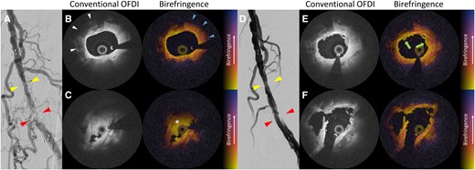
(A) Pre-angiogram. (B) Intravascular polarimetry images before excimer laser atherectomy at the proximal part of the in-stent restenosis (cross-section shown by yellow triangles in A). (C) Intravascular polarimetry images before excimer laser atherectomy at the distal part of the in-stent restenosis (cross-section shown by red triangles in A). (D) Angiogram after excimer laser atherectomy. (E) Intravascular polarimetry images after excimer laser atherectomy at the proximal part of the in-stent restenosis (cross-section shown by yellow triangles in D). (F) Intravascular polarimetry images after excimer laser atherectomy at the distal part of the in-stent restenosis (cross-section shown by red triangles in D). OFDI, optical frequency domain imaging.
After the ELA procedure, IVP images revealed a small amount of fibrin or white thrombi formation (green arrows, areas with homogeneous low birefringence in Figure 1E), while the sheet calcification and homogeneous plaques were not ablated. Given that organizing thrombi were evaporated (Figure 1F), subsequent drug-coated angioplasty was successfully performed without any distal embolization.
Excimer laser atherectomy is useful for ablating hyperplastic tissues without severe calcification.2 A pathological study demonstrated that fibrin deposition can occur due to thermal injury during ELA.3 In our case, IVP was helpful for understanding the detailed effects by ELA in vivo. Intravascular polarimetry with OFDI can provide tissue characterization of underlying plaque morphologies in great detail during endovascular intervention.
Acknowledgements
The authors are grateful to clinical engineers at our institute for their technical cooperation.
Consent: The authors confirm that written consent for the submission and publication of this case report has been obtained from the patient in line with the COPE guidance, including the use of images and associated text.
Funding: None declared.
Data availability
The data underlying this article will be shared on reasonable request to the corresponding author.
References
Author notes
Conflict of interest: None declared.

Comments