


Abstract
Coronary computed tomography angiography (CCTA) and magnetic resonance imaging (MRI) can predict periprocedural myocardial injury (PMI) after percutaneous coronary intervention (PCI). We aimed to investigate whether integrating MRI with CCTA, using the latest imaging and quantitative techniques, improves PMI prediction and to explore a potential hybrid CCTA–MRI strategy.
This prospective, multi-centre study conducted coronary atherosclerosis T1-weighted characterization MRI for patients scheduled for elective PCI for an atherosclerotic lesion detected on CCTA without prior revascularization. PMI was defined as post-PCI troponin-T > 5× the upper reference limit. Using deep learning-enabled software, volumes of total plaque, calcified plaque, non-calcified plaque (NCP), and low-attenuation plaque (LAP; < 30 Hounsfield units) were quantified on CCTA. In non-contrast T1-weighted MRI, high-intensity plaque (HIP) volume was quantified as voxels with signal intensity exceeding that of the myocardium, weighted by their respective intensities. Of the 132 lesions from 120 patients, 43 resulted in PMI. In the CCTA-only strategy, LAP volume (P = 0.012) and NCP volume (P = 0.016) were independently associated with PMI. In integrating MRI with CCTA, LAP volume (P = 0.029), and HIP volume (P = 0.024) emerged as independent predictors. MRI integration with CCTA achieved a higher C-statistic value than CCTA alone (0.880 vs. 0.738; P = 0.004). A hybrid CCTA–MRI strategy, incorporating MRI for lesions with intermediate PMI risk based on CCTA, maintained superior diagnostic accuracy over the CCTA-only strategy (0.803 vs. 0.705; P = 0.028).
Integrating MRI with CCTA improves PMI prediction compared with CCTA alone.
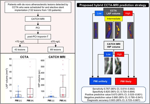
In three-dimensional views of CCTA, purple regions correspond to the outer vessel boundaries, violet regions to the lumen, orange regions to LAP and yellow regions to calcified plaque. On multi-planar reformatted images from CATCH MRI, the orange overlay indicates HIP. CATCH, coronary atherosclerosis T1-weighted characterization; CCTA, coronary computed tomography angiography; CI, confidence interval; HIP, high-intensity plaque; LAP, low-attenuation plaque; MRI, magnetic resonance imaging; PCI, percutaneous coronary intervention; PMI, periprocedural myocardial injury.


