Abstract
Chronically impacted sharp foreign bodies (FB) of the cervical or upper thoracic oesophagus can present a diagnostic dilemma. Fifty percent of sharp objects tend to lodge in the upper oesophagus and frequently cause perforation. A multidisciplinary approach is warranted. Eventually, surgery by cervical oesophagotomy/thoracotomy may be required. The Weerda diverticuloscope offers excellent endoscopic visibility and access to the lumen of the upper oesophagus allowing for removal of FBs and mitigating risks of a major surgical procedure.
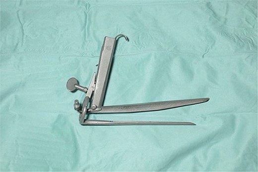
We report an interesting case of a 27 year old gentleman being investigated for dysphagia. The patient notes were obtained from the electronic patient records. The radiological images were sourced from the picture archiving and communication system (PACS) and endoscopy from stored images. The patient was radiologically diagnosed as having T4 malignancy but consecutive biopsies suggested Actinomycosis. Patient thereafter recollected having a fist fight and losing his dentures. This triggered the use of the Weerda diverticuloscope, a bivalved rigid diverticuloscope (Karl Storz, Tuttlingen, Germany). It has a distal fibreoptic illumination that is designed for trans-oral stapling of pharyngeal pouches
Under general anaesthesia, the Weerda diverticuloscope was introduced into the cervical oesophagus. The tips of the jaws positioned just proximal to the embedded dental prosthesis. The diverticuloscope was suspended for stabilisation in the same manner as for endoscopic stapling of a pharyngeal pouch and the jaws were distracted to allow endoscopic access. A 5mm rigid telescope was passed alongside a pair of heavy endoscopic scissors and the prosthesis was divided into 3 parts. The central intraluminal component was removed first using endoscopic forceps, followed by gentle disimpaction and extraction of the remaining two components embedded in the oesophageal wall.
Foreign bodies in the oesophagus rarely can mimic cancer or chronic granulomatous diseases. A multidisciplinary approach is occassionally recommended in their management. There have been a few published reports of the use of the Weerda diverticuloscope to remove upper oesophageal FBs successfully. It’s use, however, is not often considered routinely. We recommend the use of this technique at centres where expertise in endoscopic management of Zenker’s diverticula is available and when endoscopic retrieval has failed.