


Abstract
Prostaglandin analogues (PGAs; a first-line antiglaucoma treatment) have been remarketed as popular eyelash-lengthening serums due to their lash-lengthening and lash-thickening side effects. Periorbital volume loss is now a well-established side effect of topical PGAs used to treat glaucoma (prostaglandin-associated periorbitopathy) but has not, to date, been listed as a potential side effect of lash-lengthening serums containing PGAs.
The aim of this study was to identify whether periorbital fat/volume loss is seen in users of PGA lash lengtheners.
This investigation comprised a case report and an informal randomized controlled study comparing “before-and-after” color photographs displayed on the websites of manufacturers of PGA-containing lash lengtheners (PGALLs) (ie, containing bimatoprost, norbimatoprost, isopropyl cloprostenate, dechloro-dihydroxy-difluoro-ethylcloprostenolamide, or methylamido-dihydro-noralfaprostal) vs 2 control groups: non-PGALLs (NPGALL) and false eyelashes (FLs). Expert and layperson blinded graders used a purpose-designed grading system to identify subtle signs of periorbital fat/volume loss over time.
A 35-year-old female developed thin, wrinkled, darker skin, and periorbital hollowing after 10 months of treatment with Lash Boost (Rodan & Fields, San Francisco, CA), containing isopropyl cloprostenate, which reversed 6 months after discontinuation. Fifteen “before-and-after” pairs of photographs (PGALL, n = 10; NPGALL, n = 3; FL, n = 2) were graded by 5 graders (3 expert, 2 layperson). Mean grading score was 8.2 (of 19) in the PGALL group, 2.3 in the NPGALL group, and 3.2 in the FL group. PGALL scores were significantly higher than scores in the NPGALL (P < 0.001) and FL (P = 0.017) groups.
Review of commercial “before-and-after” photographs suggests that PGALL users develop changes compatible with prostaglandin-associated periorbitopathy. Consumers must be aware of the possibility of periorbital volume loss prior to commencing treatment with PGALLs. Often the customer-facing product ingredient list contains no mention of PGAs.
See the Commentary on this article .
Eyelash enhancement forms a large part of the cosmetics industry, with many options now available to those keen to improve the appearance of their eyelashes. Traditional options range from mascara to false lashes, and more recently have included lash lengtheners containing ingredients such as “proprietary peptides,” vitamins, and natural extracts.1 However, the industry was revolutionized in 2008 by the introduction a 0.03% bimatoprost ophthalmic solution, marketed under the trade name Latisse (Allergan Inc., Irvine, CA).
Bimatoprost is a prostaglandin analogue (PGA)—a family of drugs which in eye-drop form provide efficient and stable control of intraocular pressure with relatively few side effects, and are now a popular first-line antiglaucoma treatment. Many patients prescribed PGA eye drops noted lengthening, thickening, and darkening of their eyelashes.2-4 This phenomenon is believed to occur due to the interaction of prostaglandin and prostamide analogues with the prostanoid receptors found in hair follicles.5 Harnessing the potential of these, possibly favorable, side effects in the cosmetic industry, Allergan Inc. released a 0.03% bimatoprost ophthalmic solution (identical to that used in the treatment of glaucoma—Lumigan [Allergan Inc.]) under the trade name Latisse, which was licensed by the US FDA for application to the lash roots as a treatment for eyelash hypotrichosis.6,7 It should be noted that Allergan discontinued 0.03% Lumigan in 2015 due to its adverse event profile—it reported that use of the 0.01% Lumigan preparation was associated with 25% fewer overall treatment-related adverse events (vs 0.03% Lumigan), but with a similar efficacy profile.8 However, Allergan’s eyelash formulation (Latisse) continues to be provided at the higher 0.03% concentration.
PGAs created a paradigm shift in the perception of how much eyelash lengthening was possible, and as their popularity has grown many more products containing PGAs have been brought to the market, including, but not limited to: Lash Boost (Rodan & Fields, San Francisco, CA), Revitalash (Revitalash Cosmetics, Ventura, CA), Show Lash (LeVaye’ Cosmetics, Pekin, IL), Xlash (Xlash Cosmetics, Stockholm, Sweden), GrandeLASH-MD (Grande Cosmetics, Valhalla, NY), Eyelash Activating Serum (M2 Beaute, Cologne, Germany), neuLASH (Skin Research Laboratories, Moorpark, CA), and RapidLash (International Research Laboratories, Mashpee, MA)—although no other has yet required, or received, US FDA approval.
In 2004, upper eyelid sulcus deepening was reported in 3 patients being treated with topical bimatoprost for glaucoma.9 Following the reporting of many more similar cases, the clinical entity “prostaglandin-associated periorbitopathy” (PAP) was defined, variably, as the development of periorbital fat/volume loss, associated with deepening of the upper eyelid sulcus, heightening of the upper eyelid crease, ptosis, mild enophthalmos, involution of dermatochalasis, inferior scleral show, and flattening of the lower eyelid bags, due to the use of topical PGA.10-14 It has now been confirmed that these changes occur as a result of PGA-induced lipolysis and inhibition of adipogenesis.15-18 Two grading systems for PAP have been previously described: one in which unilateral grading was performed, based on color photography, with the contralateral (untreated) eye serving as a control; and one in which unilateral grading was performed based on clinical examination.14,19
Although periorbital volume loss and PAP have not, to date, been listed as potential side effects of lash lengtheners containing PGA, there has been growing anecdotal concern (particularly within online forums) that this may be the case. We describe here a case that illustrates this issue.
Case Report
A 35-year-old female presented due to concerns relating to her periocular appearance. She had started applying Lash Boost, containing isopropyl cloprostenate, 10 months previously. Over this period she had become increasingly aware that the skin around her eyes appeared thinner, dry, wrinkled, and darker, and that the periocular region looked more hollow (Figure 1). The patient found these changes very distressing, particularly as she had not been warned that they might occur. She stopped using the PGA-containing lash lengthener and was reviewed again 6 months later, at which time the changes had largely resolved. The patient reported a 90% to 95% return to what she felt was her pre-PGA appearance.
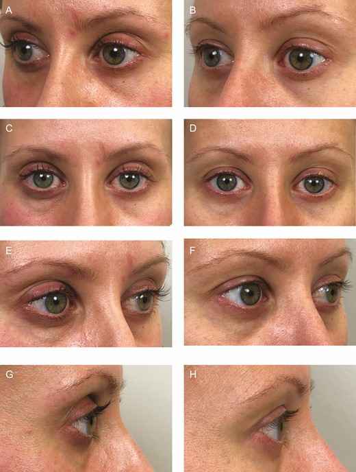
Color photographs of a 35-year-old female after taking prostaglandin analogue lash lengtheners for 10 months (A, C, E, G), and 6 months after stopping treatment (B, D, F, H). The left oblique (A, B), frontal (C, D), right oblique (E, F), and right lateral (G, H) views are presented.
Comparing the “before-and-after” photographs available from the official websites of most products, periorbital volume loss appears to be obvious to the trained eye. Based on these comparisons we hypothesize that lash lengtheners containing PGA cause periorbital fat/volume loss, albeit likely to a lesser degree than seen in patients prescribed PGA to treat glaucoma, and in designing this study, sought to identify whether these changes are present, in addition to establishing a reproducible means by which volume loss could be identified.
METHODS
An informal blinded randomized controlled study was performed based on color photographs of the users of lash-lengthening products. “Before-and-after” color photographs of lash-lengthener users were readily available from the official websites of most products (Latisse, LashBoost, Revitalash, Show Lash, GrandeLASH-MD, Eyelash Activating Serum, neuLASH, RapidLash).20-29 These images were collected between March and April 2020.
Photographs were only included if they were well-lit, included the entire periocular region, and where the user’s face was free from makeup and was held in a neutral position (in particular, not smiling). Collected photographs were added to an image bank, arranged into 3 groups: 1 surrogate “treatment group” (prostaglandin analogue lash lengthener [PGALL]), and 2 “control groups” (non–prostaglandin analogue lash lengthener [NPGALL] and false lashes [FL]).
PGALLs were identified by inspection of each product’s ingredient list. PGAs were not solely identified by the inclusion of bimatoprost. A number of other, less obvious, ingredients signaled the presence of a PGA, including: norbimatoprost, isopropyl cloprostenate, dechloro-dihydroxy-difluoro-ethylcloprostenolamide, and methylamido-dihydro-noralfaprostal.
In order to reproducibly measure the severity of fat/volume loss in the periocular region of PGALL users, a reproducible grading system was required to detect the more subtle changes expected in this cohort. The authors extensively reviewed images of the users of PGALLs, and also patients with confirmed PAP (obtained from a range of previous publications), and identified the key periocular changes which were felt to be the most significant in PGALL users. A grading system was then developed which enabled the recording of both subtle and marked changes, could be measured from only color photography (rather than clinical examination), and could be used by a layperson educated in scrutinizing the periorbital region.
The grading system is outlined in Table 1. The various periocular anatomic landmarks referenced in this grading system are illustrated in Figure 2. Grading may be either unilateral or bilateral. For the latter, the highest scoring side for each domain was used to calculate the total bilateral score. The system is anatomically organized, with features categorized to 4 main periocular regions: upper lid; lower lid; medial canthus; lateral canthus (Figure 2A,B). For each domain, 1 point was given for each feature present (to a maximum of 2 points), except in the instance of skeletonization (or reveal) of the bony orbital rim, which in all cases resulted in a score of 2 points. A final total score was calculated, from a total of 19 total available points.
Periorbital Fat/Volume Loss Grading System
| Region | Score 0 = no change between photographs, 1 = only 1 change noted in this region, 2 = more than 1 change noted in this region, or presence of a “grade 2” feature |
|---|---|
| Upper lid 1. Preseptal region (ie, central sub-brow region) - Volume loss: preseptal (or central sub-brow) skin appears less full, hollowing of the superior sulcus - Associated increase in skin fold height - Associated increase in lid show | 0 1 2 |
| 2. Pretarsal region - Volume loss: pretarsal skin appears less full - Flattening of the convex curvature of the pretarsal skin | 0 1 2 |
| 3. Lateral sub-brow region - Volume loss: flattening of the normal convex contour, appears less full - Increased reveal/skeletonization of the superior/superolateral orbital rim (grade 2, even if only feature present) | 0 1 2 |
| 4. Brow - Descent of the brow (either of the entire brow or portions of the brow) | 0 1 - |
| Lower lid 5. Pretarsal region - Volume loss: pretarsal skin appears less full - Flattening of the convex curvature of the pretarsal skin (ie, above the inferior border of the tarsal plate) - Increased angulation/deepening of the lower eyelid crease hollow or septal confluence hollow | 0 1 2 |
| 6. Tear-trough - Volume loss: hollowing or deepening of the tear trough - Medial fat pad loss | 0 1 2 |
| 7. Lid-cheek junction/palpebromalar junction - Flattening of natural convexity ± development of concavity | 0 1 - |
| 8. Lower lid height - Increase in inferior scleral show | 0 1 - |
| 9. Rhytids - Increase in area of skin with evident rhytids, number of rhytids, or depth of rhytids | 0 1 - |
| Medial canthus 10. Medial canthal tendon - Increased reveal of medial canthal tendon due to surrounding volume loss | 0 1 - |
| Lateral canthus 11. Orbital rim - Increased reveal/skeletoniation of the lateral orbital rim (Grade 2) | 0 - 2 |
| 12. Lateral raphe region - Volume loss in lateral raphe region - Increase in area of skin with evident rhytids, number of rhytids, or depth of rhytids | 0 1 2 |
| Total | Score = __________ out of 19 |

Periocular anatomic landmarks shown on a 35-year-old female: (A) “creases and hollows”; (B) other landmarks referenced in the grading system.
Grading of the image bank was performed by 5 blinded graders. Of these, 3 were ophthalmologists with specialist training in oculoplastic surgery, considered to be expert graders, all of whom are authors (A.J., L.O., K.U.). In addition, 2 laypeople, following brief training in scrutinization of the periorbital region, also graded the image bank in order to identify whether any detected differences between the study groups were also perceivable to the “untrained eye.”
Based upon the difference in lash length, all graders knew that the pairs of images represented “before and after” the use of a product, but were masked to whether the product was a PGALL, an NPGALL, or FLs. Each grader was masked to the findings of other graders, and entered their gradings into a separate electronic database. Grading was then repeated by each grader at an interval of 2 days in order to allow the evaluation of intrarater reliability. As this study involved no “real” participants, the NHS Health Research Authority deemed that ethical approval was not required.
Statistical Analysis
Krippendorff’s α was used to examine both inter- and intrarater reliability for the total PAP scores.30 Krippendorff’s α is preferred to weighted (and unweighted) Cohen’s κ as the PAP scores obtained are interval numeric variables, with increasing scores representing increasing PAP severity. Krippendorff’s α also allows the analysis of interrater reliability where there are more than 2 graders. Interrater reliability was analyzed based on each grader’s second grading.
Statistical analysis was performed with the statistical software Real Statistics (via Microsoft Excel). Landis and Koch’s classification of κ values, which may be applied to the result (α) of a Krippendorff’s α analysis, was used to describe the strength of agreement: slight (≤0.20), fair (0.21-0.40), moderate (0.41-0.60), substantial (0.61-0.80), and almost perfect (0.81-1.00).31,32
Mann-Whitney U tests were used to compare the grading scores between groups. As described in the Results section, low sample size in the layperson gradings for the FL group prevented use of the Mann-Whitney U test, and a parametric test (the 2-tailed independent t test) was used instead.
RESULTS
Thirty “before-and-after” pairs of photographs of women who had used PGALLs were obtained. Ten of these passed an image-quality check, as described above, and were included in the PGALL group. There were a further 3 image pairs in the NPGALL group, and 2 in the FL group. None of the image pairs were of male users. It was not possible to ascertain the age of the imaged product users.
Grading System Validation
Intrarater (or test-retest) reliability was 0.979 (“almost perfect” agreement) for grader 1, 0.847 (“almost perfect” agreement) for grader 2, and 0.781 (“substantial” agreement) for grader 3. Interrater and intrarater reliability measures are outlined in Table 2. Based on their second grading attempts, agreement between the 3 expert graders was 0.545 according to Krippendorff’s α method. Based on Landis and Koch’s categorizations of agreement, this represents a “moderate” agreement, and is consistent with the inherently subjective nature of discerning subtle changes in the periorbital region. The second gradings of each grader are illustrated in Figure 3.
Interrater and Intrarater Reliability for the Grading Scale
| Test | Ratings analyzed | α estimate | Level of agreement |
|---|---|---|---|
| Interrater reliability | Grader 1 (second), grader 2 (second), grader 3 (second) | 0.545 | Moderate |
| Intrarater reliability | Grader 1 (first), grader 1 (second) | 0.979 | Almost perfect |
| Grader 2 (first), grader 2 (second) | 0.847 | Almost perfect | |
| Grader 3 (first), grader 3 (second) | 0.781 | Substantial |
![Grading scores for each grader, based on their second grading. The images, which were randomized during grading, have been reorganized into the 3 study groups (prostaglandin analogue lash lengtheners [PGALL], non–prostaglandin analogue lash lengtheners [NPGALL], and false lashes [FL]) for illustrative purposes.](https://oup.silverchair-cdn.com/oup/backfile/Content_public/Journal/asj/42/11/10.1093_asj_sjac156/1/m_sjac156f0003.jpeg?Expires=1747861109&Signature=chtnWoKoLvfVhBJAmG9h14~JxUG6QpBgcBpVxK3XXYHKwiLCsq7GE-aNjQAkeYyPSTZ7K2S8hOiPBZkwUE~eAoZlobuEAzanpo6ylQoeOeayU7cfP2h2kcgMEwTmsjYhCojqsHYnokv1RXefNGuZ3CuH8XOgzUkl5szTbwmwa-v9DA-lsrpr2hvFFZ3aKKJCzopKW-vtRr50ea8GQRvFgDCf3j80E6hoz-3QHDHevVOTBEtTRn9napn72A0cm7jIVqU8qfzIMs1oSVEemHXVPwZP78tTD6inLPZbaPLdC~~0xB~PwVkzxX69-Yh~pLDj0X5s8uXu~LXZOOUUjy2xZQ__&Key-Pair-Id=APKAIE5G5CRDK6RD3PGA)
Grading scores for each grader, based on their second grading. The images, which were randomized during grading, have been reorganized into the 3 study groups (prostaglandin analogue lash lengtheners [PGALL], non–prostaglandin analogue lash lengtheners [NPGALL], and false lashes [FL]) for illustrative purposes.
Results of Grading
Expert Graders
Analysis is based on each grader’s second grading attempt. Expert graders reported the greatest fat/volume loss in the PGALL group, with similar lower levels of loss in the NPGALL and FL groups. The mean (median) grading score was 8.2 (7.5) in the PGALL group, 2.3 (1.0) in the NPGALL group, and 3.2 (2.0) in the FL group. Five number summaries for each group are illustrated in Figure 4.
![Box-and-whisker plots illustrating the 5 number summaries (minimum, Q1, median, Q3, maximum) for each group (prostaglandin analogue lash lengtheners—PGALL [n = 10], non–prostaglandin analogue lash lengtheners—NPGALL [n = 3], false lashes—FL [n = 2]), once again based on each expert grader’s second attempt. Corresponding t test outputs and P values are also illustrated.](https://oup.silverchair-cdn.com/oup/backfile/Content_public/Journal/asj/42/11/10.1093_asj_sjac156/1/m_sjac156f0004.jpeg?Expires=1747861109&Signature=QxnmlkU8U9lnJ7KMxAjQGsc~2TNk1L-nVlS2l4VYSDi0mfEZFqzCL04HU5h4AFC7anqFEmvofDe2rB00LPxm6SvM5rLdOK2YpZhdF~K41EdAGtu0NNPp1MoSv7T0nRXOvojnS-brXbQUrAajx1VutU8NbewKo8dgHhF-JjKBwBSQFCghaRA9qR4lIRKGleEz3ZAEhWEaMqc7m0V2FDx52eJfras5nZVUJDCqU83s7qKokZw9khzCWrSJvsVYOOQOqt4GNCUq8muvMRlEcv9O8Zr~QKWhKk~ketbILCciC4d0mdo~EM~Qi6I0kUM9QCLzUUShB~6Vd3nJiTf0loXHBA__&Key-Pair-Id=APKAIE5G5CRDK6RD3PGA)
Box-and-whisker plots illustrating the 5 number summaries (minimum, Q1, median, Q3, maximum) for each group (prostaglandin analogue lash lengtheners—PGALL [n = 10], non–prostaglandin analogue lash lengtheners—NPGALL [n = 3], false lashes—FL [n = 2]), once again based on each expert grader’s second attempt. Corresponding t test outputs and P values are also illustrated.
A Mann-Whitney U test demonstrated that grading score was significantly higher in the PGALL group than in the NPGALL “control” group (U = 39.5, P = 0.002). Grading score was also significantly higher in the PGALL group than in the FL “control” group (U = 35, P = 0.021). No statistically significant difference was identified between the 2 “control” groups (NPGALL and FL) (U = 24, P = 0.772).
The grading scores for PGALL users were then considered by region. The 3 domains with the highest mean score (ie, the regions with the most marked volume loss identified) were the upper lid preseptal/sub-brow region (1.20, out a maximum total of 2.0), upper lid pretarsal region (1.20) and lower lid pretarsal region (1.13). These regions are demonstrated in Figure 5.
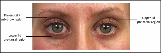
Color photograph of reported case of a 35-year-old female (after 10 months of PGALL treatment) used as an example image to illustrate the 3 regions with most marked volume loss in this study. Highest mean scores were: 1.20 (out a maximum total of 2.0) in the upper lid preseptal/sub-brow region; upper lid pretarsal region (1.20); and lower lid pretarsal region (1.13). To demonstrate the use of the outlined grading system (Table 1) with our reported case (Figure 1), readers might assume that this female patient’s appearance before starting a PGALL was similar to that seen in Figure 1D (frontal view, which was actually taken 6 months after stopping PGALL, when the patient reported a 90% to 95% resolution of her periocular changes). By looking at this image alongside Figure 1C (frontal view, taken after 10 months of PGALL treatment) we can construct an ad hoc “before-and-after” image pair, which we can grade (for demonstration purposes only) for periorbital fat/volume loss. In this case, the image pair would have been given a grading score of 6 (out of 19), as outlined. Upper lid = 3: preseptal = 2 (preseptal/central sub-brow skin appears less full, and there is hollowing of the superior sulcus, associated with increased skin fold height and an increase in lid show); pretarsal = 1 (pretarsal skin appears less full); lateral sub-brow region = 0 (no significant change); brow = 0 (no significant change). Lower lid = 2: pretarsal = 1 (pretarsal skin appears less full); tear trough = 0 (no significant change); lid-cheek junction = 0 (no significant change); lower lid height = 0 (no significant change); rhytids = 1 (increase in number of evident rhytids). Medial canthus = 1: medial canthal tendon = 1 (increased reveal of medial canthal tendon). Lateral canthus: orbital rim = 0 (no significant change); lateral raphe region = 0 (no significant change). PGALL, prostaglandin analogue lash lengthener.
Layperson Graders
Analysis is based on each grader’s only grading attempt. The mean (median) grading score was 5.1 (4.5) in the PGALL group, 1.0 (0.5) in the NPGALL group, and 1.5 (1.5) in the FL group. Five number summaries for each group are illustrated in Figure 6.
![Box-and-whisker plots illustrating the 5 number summaries (minimum, Q1, median, Q3, maximum) for each group (prostaglandin analogue lash lengtheners—PGALL [n = 10], non-prostaglandin analogue lash lengtheners—NPGALL [n = 3], false lashes—FL [n = 2]), based on each layperson grader’s only attempt. Corresponding t test outputs and P values are also illustrated.](https://oup.silverchair-cdn.com/oup/backfile/Content_public/Journal/asj/42/11/10.1093_asj_sjac156/1/m_sjac156f0006.jpeg?Expires=1747861109&Signature=CX47E5dFyGfqh18KyHNmyKJMWfH~w8eQGMIeMLhBJcdlKS19eWNr6YUjyfnIxOl~60X6kLMQ3xUfhHQkJ1bgtuspdXPpvoPCWEy1ngjTV1AFpJuXA1RJuvBvpDXpJAHF5bJbwa4~M574VAn7VUxrHSAMSCTCx-yuBl6aVqsr76E8kB1QbJiYqITTCrhPblpF5Fcwr6K45C8tkjf4Me-orfMQJ37Ew1U~cz6vah-UFFY8BgVgJrYLwjWYYKFUW2GnnpMZiv9VwCQIjJ6eV1XiLH190sDsX9rJVjgQvAK~zbS6Gq~SLFhRrLDw8jngGS~zGXBG2jrD52qEXr3NpNP9hA__&Key-Pair-Id=APKAIE5G5CRDK6RD3PGA)
Box-and-whisker plots illustrating the 5 number summaries (minimum, Q1, median, Q3, maximum) for each group (prostaglandin analogue lash lengtheners—PGALL [n = 10], non-prostaglandin analogue lash lengtheners—NPGALL [n = 3], false lashes—FL [n = 2]), based on each layperson grader’s only attempt. Corresponding t test outputs and P values are also illustrated.
Mann-Whitney U tests cannot be performed with sample sizes of 5 or fewer. Given that the FL group had only 4 measurements (2 image pairs × 2 layperson graders), the Mann-Whitney U test could only be used to compare the PGALL and NPGALL groups. Grading score was significantly higher in the PGALL group than in the NPGALL “control” group (U = 12.5, P = 0.007).
Parametric tests (a 2-tailed independent t test) were used compare the remaining pairs of groups. Grading score was also significantly higher in the PGALL group than in the FL “control” group (t(22) = 2.51, P = 0.046). No statistically significant difference was identified between the 2 “control” groups (NPGALL and FL) (t(8) = –0.49, P = 0.560).
DISCUSSION
The development of periorbital fat/volume loss, although widely acknowledged in patients taking topical PGAs to treat glaucoma, has not previously been described in the context of lash lengtheners containing PGAs. That being said, numerous accounts of similar periocular changes have been self-reported by the users of such products on the internet, in a range of forums, blogs, YouTube videos, and online articles. In this study, periorbital volume loss was detected in all patients taking PGALLs, and was most noticeable in the upper lid preseptal region and the upper and lower lid pretarsal regions. Although periorbital volume loss is usually considered to be a negative—the loss of the youthful fullness of the periocular region—it is possible that some may feel this to be a positive. However, the authors believe that it is still important to highlight this side effect to both the cosmetic industry and its customers.
The process of compiling a suitable study image bank also identified a further issue worthy of highlighting. In numerous cases it was not immediately obvious that a lash lengthener contained PGAs. Often the customer-facing ingredient list contained no mention of a PGA, and it was only identified on careful interrogation of the separate product information sheet. Greater clarity is certainly needed in such products. Concerns that “over-the-counter” PGA lash lengtheners have not been studied for safety and efficacy, nor undergone a comprehensive pharmacovigilance program, have previously been raised.1 Importantly, whereas patients with glaucoma starting a PGA eye drop will have a discussion with the prescribing doctor regarding the side-effect profile, consumers “self-prescribing” PGA lash lengtheners, or being offered them by their beautician/aesthetician, will not have this information made available to them.
A particular strength of this study is that the images analyzed were those that had been obtained directly from the websites of the companies that market PGA lash lengtheners, and could be viewed as a representation of the ideal outcome. These images are readily accessible on the official websites of most PGA lash lengtheners.
A notable feature of this study is the development of a novel periorbital fat/volume loss grading system which may be used both by the prescribers of PGALLs, and the users themselves. Validation of this grading system was undertaken, which demonstrated “moderate” interrater reliability and “almost perfect” or “substantial” intrarater reliability in the hands of expert graders. The inclusion of gradings made by laypeople, although not validated by reliability analysis, highlights that the development of volume loss following PGALL use is noticeable not just to the trained eye, but also to the users themselves, and their friends and relatives. Furthermore, the users of PGALLs could potentially utilize this grading system to assess their own photographs for the development of periorbital fat/volume loss.
Our study contained smaller numbers of image pairs within both “control” groups, in large part due to the fact that image quality for these products was variable and the women pictured were often wearing makeup. As such, fewer of these images met the initial quality-control check. Despite this quality check, there will remain some variation in lighting intensity and direction, magnification, head position, and facial expression. The likelihood that some of the images may have been Photoshopped for improved appearance was also considered, even though this would not be easily recognizable when examining any one particular image. Despite small numbers in these groups, and the possibility that these images had been Photoshopped for improved appearance, statistical analysis was able to detect significant differences in grading scores. However, our graders reported a small degree of volume loss even in the FL group, which should not be present. This is likely a result of the smaller numbers of image pairs in this group, in addition to the inherent bias of asking graders to look for volume loss in an “after” photograph.
A further limitation of this study is that many of the features characteristic of periorbital fat/volume loss are not specific to PAP or PGALL use. In particular, the effects of aging on the periocular region are hard to discriminate from PAP, and in fact, early reports of PGA-associated periocular changes were often dismissed as due to aging.10,14 The potential for aging-related periocular change to confound the results of our study is heightened by the fact that grading was based on 2 photographs taken at different time points, the time between which was not always known. Further prospective study of periorbital fat/volume loss in PGALL users could control for aging-related change by grading both eyes simultaneously after unilateral PGALL use, although the cosmetic implications of this are not likely to be tolerated by study participants.
Finally, the grading system outlined in this study aims to evaluate fat/volume loss from color photographs. It should be highlighted that such a grading system can only measure surface changes (such as skin folds, and perceived volume loss) which are believed to reflect the degree of underlying volume loss.
CONCLUSIONS
Our study suggests that the users of PGA lash lengtheners develop periorbital fat loss, which is particularly evident in the upper lid preseptal region and the pretarsal region of both lids. This study provides the proof of concept, and an appropriate grading tool, to perform a prospective study to confirm these findings. It is important that clinicians and consumers are made aware of the possibility of periorbital volume loss prior to commencing treatment with PGALLs.
Disclosures
The authors declared no potential conflicts of interest with respect to the research, authorship, and publication of this article.
Funding
The authors received no financial support for the research, authorship, and publication of this article.

